Clear Sky Science · en
Impact of thermal and physiological denoising on laminar functional connectivity
Why cleaning up brain scans matters
Modern brain scanners can now peer into the six thin layers of the human cortex, letting scientists ask not just which region is active, but which depth within that region is sending or receiving information. Yet these ultra-detailed images are full of different kinds of “noise” from the scanner, the blood vessels, and even the heartbeat and breathing of the person in the machine. This study asks a practical question with big implications: if we carefully clean these noisy signals, can we get a truer picture of how activity travels between layers in a key movement area of the brain?
Looking at layers inside the movement area
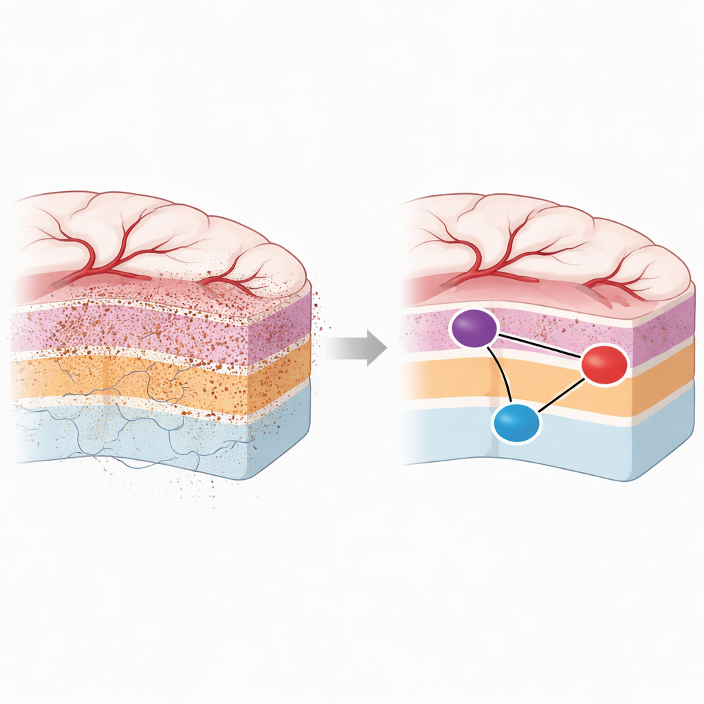
The researchers focused on the primary motor cortex, the strip of brain tissue that helps control voluntary movements, especially of the hand. This region, like the rest of the cortex, is built from six stacked layers that differ in how they receive and send information. Upper layers tend to receive inputs from other areas, while deeper layers carry outputs to other brain regions and to the spinal cord. Using a very strong 7-tesla MRI scanner and tiny voxels less than a millimeter wide, the team recorded spontaneous (resting-state) activity from the hand area of motor cortex and from neighboring somatosensory and premotor regions that exchange signals with it.
The problem of noisy and biased signals
At such fine resolution, the useful signal in these scans competes with several unwanted sources. Random “thermal” noise comes from the electronics of the scanner itself, and is especially troublesome in deeper layers where the signal is weaker. Physiological noise, in contrast, comes from the subject’s body: changes in breathing, heartbeats, and blood oxygen in large veins near the cortical surface. Because standard fMRI emphasizes signals from big veins, superficial layers can appear more active and more connected than they really are, even if those fluctuations are just vascular ripples rather than true neural communication. Without careful correction, researchers risk misreading these superficial fluctuations as strong top-layer connections between brain areas.
Testing ways to clean up the data
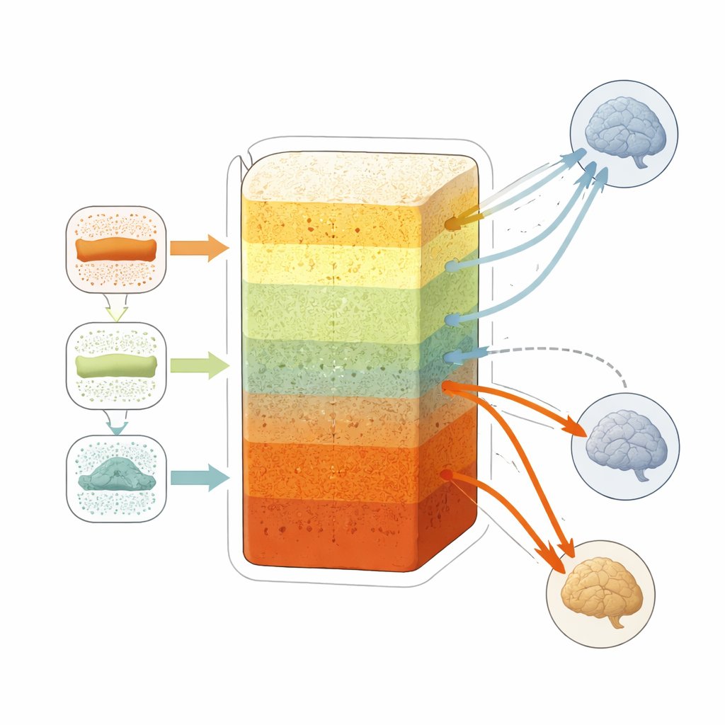
To tackle these issues, the team compared several established “denoising” steps. First, they applied an algorithm called NORDIC that is designed to suppress thermal noise in the images. Then they added motion correction, followed by one of two physiological-cleaning strategies. One, known as RETROICOR, uses recordings of the subject’s breathing and pulse to subtract out related fluctuations. The other, called aCompCor, extracts noise patterns from regions dominated by fluid or white matter inside the MRI images themselves and regresses those patterns out. By combining these steps in different ways, the researchers asked how much each method reduced unwanted fluctuations and how it altered the apparent strength of layer-specific connections between motor cortex and its neighbors.
What changed after denoising
The investigators examined several measures of data quality layer by layer, including how strongly the signal fluctuated over time and how power was distributed across different frequency bands. NORDIC had the largest overall impact, especially in deeper layers, reducing random variation and making the resting signals more stable without changing the average signal level. Physiological denoising, particularly aCompCor, had its greatest effect in the upper layers, where large veins and physiological rhythms dominate. When the team looked at functional connectivity—how tightly the activity in one region tracked that in another—they found that thermal denoising initially boosted apparent connectivity everywhere, while aCompCor then selectively trimmed away spurious upper-layer correlations, especially those involving premotor cortex and a control area that should not be strongly linked.
A clearer picture of how layers talk
After the full pipeline of thermal and physiological denoising, the resulting pattern of connections fit better with what is known from anatomy and prior high-precision studies. Upper layers of the primary motor cortex still showed stronger coupling with the neighboring somatosensory area, consistent with rich incoming sensory input to those depths. However, the earlier bias toward unusually strong upper-layer connections with premotor cortex was reduced, and signals from deeper layers became relatively more informative. In everyday terms, the study shows that careful cleaning of high-resolution brain scans can strip away misleading echoes from blood vessels and body rhythms, allowing a closer look at the true dialogue between different layers of the cortex. This makes laminar fMRI a more reliable tool for tracing the direction of information flow in the human brain.
Citation: Guidi, M., Giulietti, G., Sharoh, D. et al. Impact of thermal and physiological denoising on laminar functional connectivity. Sci Rep 16, 8602 (2026). https://doi.org/10.1038/s41598-026-37599-4
Keywords: laminar fMRI, functional connectivity, brain imaging noise, motor cortex layers, denoising methods