Clear Sky Science · en
Multimodal magnetic resonance imaging of the vastus medialis for quantitative diagnosis and grading in early-stage knee osteoarthritis and its correlation with severity
Why Your Aching Knees Start with a Thigh Muscle
Knee osteoarthritis is often blamed on worn-out cartilage and aging joints. But this study suggests that, long before X‑rays look alarming, a key thigh muscle on the inner side of the leg—the vastus medialis—quietly begins to change. By using advanced MRI techniques, the researchers show that subtle shifts in water movement, blood flow, and fat inside this muscle can help detect and grade early knee osteoarthritis, potentially opening the door to earlier, more targeted treatment.
A Common Problem, Caught Earlier
Knee osteoarthritis is one of the world’s leading causes of disability, especially in middle‑aged and older adults. Traditionally, doctors diagnose it using X‑rays that show joint space narrowing and bone spurs, along with symptoms like pain and stiffness. But by the time those changes are obvious, joint damage is often well underway. New Chinese guidelines for early-stage knee osteoarthritis broaden the definition to include milder X‑ray findings and subtle MRI signs in cartilage and bone. Even so, those criteria still depend heavily on the eye and experience of the radiologist and can miss very early disease or mislabel normal knees as diseased.
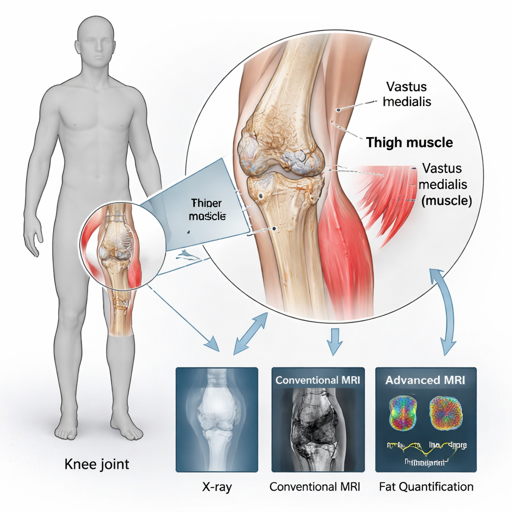
The Muscle that Guards the Knee
Growing evidence points to the muscles around the knee—especially the quadriceps on the front of the thigh—as major players in joint health. Within this group, the vastus medialis, located on the inner side of the thigh, is particularly important for stabilizing the kneecap and balancing forces across the joint. Studies show that this muscle is often the first to weaken, shrink, and fill with fat in people with knee problems. Fatty, weakened muscle not only reduces support for the joint but also releases inflammatory substances that can worsen cartilage damage and pain. The authors reasoned that if they could measure these early changes inside the vastus medialis precisely, they might gain a more sensitive window into the earliest stages of knee osteoarthritis.
Looking Inside Muscle with Advanced MRI
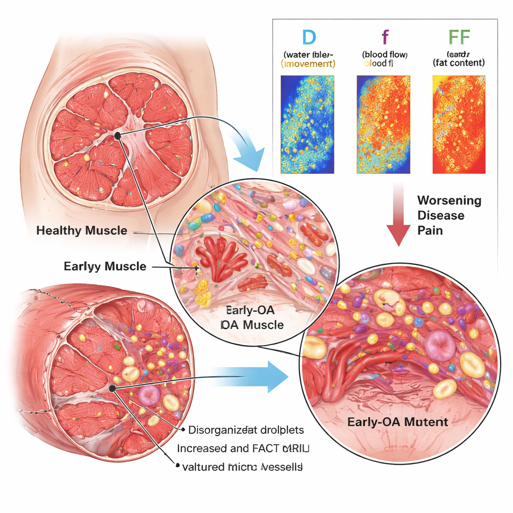
The team studied 207 knees from 168 people aged 40–70, comparing healthy volunteers with patients who met criteria for early-stage knee osteoarthritis. All participants underwent standard X‑rays and conventional knee MRI, plus two specialized MRI scans focused on the vastus medialis. One scan, called intravoxel incoherent motion diffusion‑weighted imaging (IVIM‑DWI), tracks how water molecules move in tissue and how blood flows through tiny vessels. From this, the researchers extracted three numbers: D (overall water movement), D* (a measure related to micro‑blood‑flow speed), and f (the fraction of signal coming from blood). The second scan, the fat analysis calculation technique (FACT), separates water and fat signals and yields a “fat fraction” (FF), indicating how much of the muscle volume is fat rather than muscle fibers.
What Changes with Early Knee Disease
Compared with healthy controls, people with early knee osteoarthritis showed higher D and D* values and a higher fat fraction in the vastus medialis, but a lower f value. In simple terms, water moved more freely (suggesting inflammation and tissue breakdown), micro‑blood‑flow speed appeared increased, yet the overall volume of tiny blood vessels was reduced, and the muscle contained more fat. When the researchers grouped patients by standard X‑ray severity (Kellgren–Lawrence grades 0, I, and II), D and fat fraction rose steadily from the mildest to the more advanced early grades, while the perfusion fraction f tended to fall, with the largest drop at Grade II. Importantly, higher D and fat fraction were moderately linked not only to worse X‑ray grades but also to higher pain scores on the visual analog scale, whereas f showed a weak negative link to both. D and FF also proved more stable and reliable than D*, making them especially promising as practical markers.
From Imaging Numbers to Real‑World Care
To a non‑specialist, these MRI parameters can be thought of as early warning gauges inside the thigh muscle: rising water movement (D) and fat content (FF), along with shrinking blood‑flow fraction (f), signal that the vastus medialis is inflamed, infiltrated by fat, and losing quality even while standard scans may look nearly normal. Because these muscle changes track both structural knee damage and the patient’s pain, they could help doctors diagnose early knee osteoarthritis more accurately, sort patients into stages that better match their underlying biology, and tailor non‑surgical treatments such as targeted strengthening, weight management, and anti‑inflammatory strategies. In short, by listening closely to what a single, small thigh muscle is telling us, clinicians may be able to protect aging knees before the damage becomes irreversible.
Citation: Liu, Y., Tian, D., Guo, Y. et al. Multimodal magnetic resonance imaging of the vastus medialis for quantitative diagnosis and grading in early-stage knee osteoarthritis and its correlation with severity. Sci Rep 16, 6237 (2026). https://doi.org/10.1038/s41598-026-37567-y
Keywords: early knee osteoarthritis, vastus medialis muscle, advanced knee MRI, muscle fat infiltration, knee pain imaging