Clear Sky Science · en
Design, fabrication and characterization of metamaterial absorber for sensing applications
Why this tiny surface matters
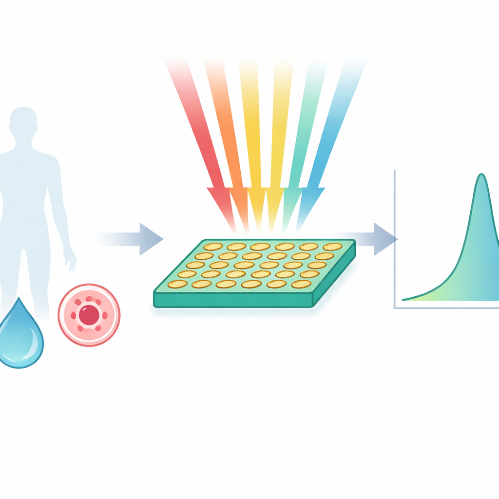
Imagine a flat, postage‑stamp‑sized surface that can tell healthy cells from cancer cells simply by how they bend invisible waves. This study presents just such a device: a specially engineered “metamaterial” surface that almost perfectly absorbs millimeter‑wave radiation and turns tiny changes in nearby biological tissue into clear, measurable signals. It promises faster, cheaper, and less invasive ways to sense disease and to monitor fluids and materials—without needing labels, dyes, or bulky lab equipment.
Building an unusual wave‑eating surface
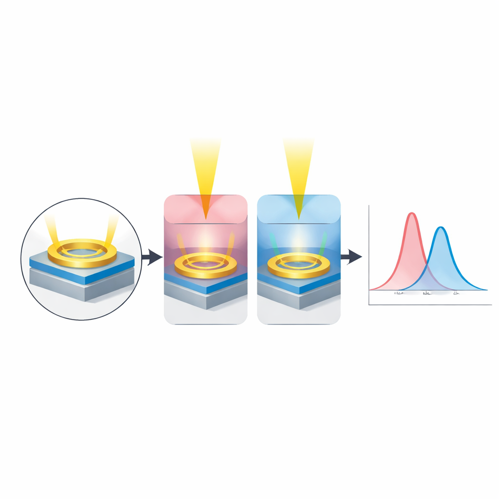
At the heart of the work is a perfect metamaterial absorber, a man‑made structure whose properties do not exist in ordinary materials. The researchers pattern two thin copper rings and connecting strips on a common circuit‑board material (FR‑4) and place a solid copper layer underneath. When millimeter‑wave radiation hits this sandwich at around 28 gigahertz—near frequencies being explored for 5G wireless—the geometry forces electric and magnetic vibrations to occur together. The bottom copper sheet blocks transmission, while the patterned top layer is carefully tuned so its effective electrical properties match those of empty space. Under these conditions, reflection almost vanishes and nearly all incoming energy is soaked up at one very sharp frequency.
From design on screen to real‑world hardware
The team first used full 3‑D electromagnetic simulations to refine the tiny dimensions of the rings and gaps so that the absorber would show a single, extremely narrow absorption peak. In the virtual model, the structure captured 99.33% of incoming radiation at 28.146 gigahertz, with the energy confined to a small region around the copper pattern. The sharpness of this peak, described by a high "quality factor," means that even slight shifts in frequency are easy to spot. To confirm the design, the researchers fabricated a 10‑by‑10 array of these unit cells on a 15‑centimeter‑square board using standard photolithography. Laboratory measurements with a horn antenna and a vector network analyzer showed a real absorption of 96.5% at 28.12 gigahertz, in close agreement with the simulations.
Turning absorption into a sensitive detector
Because the resonant frequency depends on the refractive index—how strongly a material slows and bends electromagnetic waves—the absorber can act as a sensor. The authors placed a thin layer of test material directly on top of the patterned copper. When they changed the refractive index in their simulations by only 0.05 (for example, from 1.30 to 1.35, typical of many biological fluids), the resonance shifted measurably, yielding a very high simulated sensitivity and a figure of merit that surpasses most similar sensors reported in the microwave range. Experiments using water as the test layer showed that switching from air to water moved the resonance down from about 28 to 23.5 gigahertz, still with strong absorption, confirming that the device responds robustly to realistic samples.
Spotting cancer through subtle optical fingerprints
Cancer cells often contain more protein and other dense components than normal cells, giving them slightly higher refractive indices. The researchers exploited this fact by modeling how their sensor would respond to different cell types applied as a thin layer on the metamaterial. For basal, breast, cervical (HeLa), Jurkat (a blood cancer line), MCF‑7 (breast), and PC12 (nerve‑like) cells, they compared the predicted resonance for normal versus cancerous states. In every case, the peak frequency shifted by a small but clear amount when moving from normal to cancer cells, corresponding to average sensitivities on the order of nine gigahertz per unit change in refractive index—enough to distinguish cell states without needing labels or staining.
How a small shift reveals a big change
Behind this behavior lies a simple principle similar to a tuning fork. The patterned copper rings and gaps act like tiny resonant circuits made of inductors and capacitors. Adding a sample on top changes how electric fields concentrate in the gaps, effectively altering this microscopic “spring and mass” system. A denser, higher‑index layer—such as cancerous tissue—changes the balance, shifting the pitch of the resonance. Because the metamaterial’s response is so sharply defined, these shifts stand out clearly against the background, enabling precise measurements even when the absolute changes in refractive index are small. The authors conclude that their compact, low‑cost, and highly selective absorber is a strong candidate for future sensors in high‑frequency biosensing, including early cancer detection and advanced diagnostics compatible with emerging wireless technologies.
Citation: Helaly, D.M.M., Hameed, M.F.O., Areed, N.F.F. et al. Design, fabrication and characterization of metamaterial absorber for sensing applications. Sci Rep 16, 8268 (2026). https://doi.org/10.1038/s41598-026-37524-9
Keywords: metamaterial biosensor, millimeter wave sensing, perfect absorber, cancer cell detection, refractive index sensor