Clear Sky Science · en
Plant-mediated synthesis of silver nanoparticles using Alcea rosea leaf aqueous extract and evaluation of the biological activities
Turning Garden Flowers into Tiny Medical Tools
Imagine if the leaves of a common garden flower could help fight harmful germs and even cancer cells, without relying on harsh industrial chemicals. This study explores exactly that: using the hollyhock plant (Alcea rosea) to produce ultra‑small silver particles in a cleaner, more sustainable way. The work shows how plant chemistry can reshape silver into nanoparticles and how these tiny particles behave against bacteria, free radicals, and cancer cells in the lab.
Why Silver Needs a Green Makeover
Silver has been prized for centuries for its ability to keep things clean and germ‑free. When silver is broken down into nanoparticles thousands of times smaller than the width of a human hair, its properties become even more powerful and versatile, with uses in electronics, coatings, medicines, and disinfectants. But the usual ways of making these nanoparticles often involve toxic substances, high energy use, and complicated clean‑up steps. Researchers are therefore searching for “green” routes that replace industrial chemicals with natural helpers—such as plant extracts rich in sugars, antioxidants, and other active compounds—to both form and stabilize these tiny particles.
A Medicinal Flower Does Double Duty
Alcea rosea, better known as hollyhock, is grown worldwide for its large colorful blooms and has a long history in traditional remedies for infections, inflammation, and digestive problems. In this study, scientists collected hollyhock leaves from western Nepal and prepared a simple water extract by gently heating ground leaf powder in warm water. The natural substances in this extract—flavonoids, alkaloids, and other plant metabolites—are able to donate electrons and cling to surfaces, making them ideal “kitchen chemistry” tools. When the greenish leaf extract was mixed with a solution of silver salt and the acidity was carefully adjusted, the liquid turned a deep brown, signaling that silver ions had been transformed into solid silver nanoparticles coated by plant molecules. 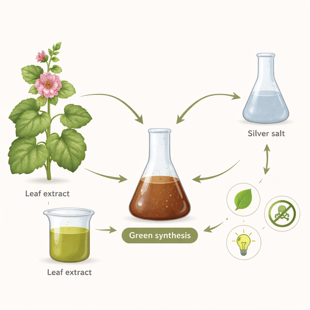
Seeing and Measuring the Invisible
To confirm what they had made, the team used several standard techniques that reveal different aspects of the particles. Light‑absorption measurements showed a sharp signal at a wavelength typical for silver nanoparticles, indicating that the metal had taken on its new nano‑form. Infrared analysis compared the plain leaf extract with the final particles and showed that groups such as oxygen‑ and nitrogen‑containing bonds had shifted, evidence that plant compounds were binding to the silver surface. X‑ray diffraction patterns revealed that the particles had a well‑ordered crystal structure, with individual crystal domains only about five nanometers across, while high‑resolution electron microscopy images showed mostly spherical clumps about 22–64 nanometers in overall diameter. Additional tests of emitted X‑rays confirmed that the material was predominantly silver, along with carbon and oxygen from the plant coating.
How the Tiny Particles Behave in the Lab
Once the particles were well characterized, the researchers tested how they performed in several biological settings. In an antioxidant test that measures how well a substance can neutralize a stable free radical, the silver nanoparticles did show protective activity, but they were much weaker than a pure plant antioxidant used as a reference. Antibacterial tests told a more promising story: the particles slowed the growth of four disease‑related bacteria, with moderate effects against Staphylococcus aureus and Shigella sonnei. However, the amount needed to fully stop and then kill these microbes was higher than that required for a standard antibiotic, suggesting that these particles are helpful but not yet strong replacements on their own. 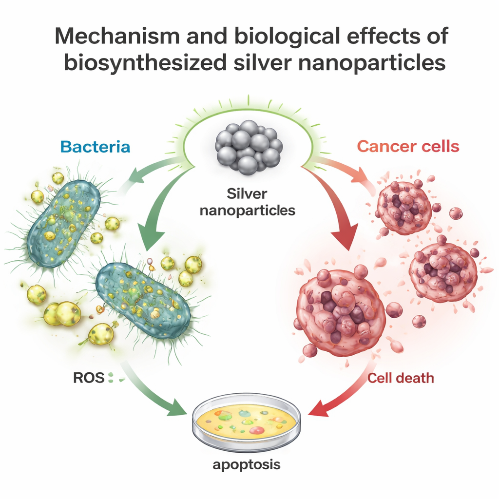
Early Hints of Anticancer Potential
The most striking results appeared when the team exposed human cancer cell lines—one from lung tissue and one from cervical tissue—to increasing doses of the hollyhock‑based silver nanoparticles. Over two days, the particles reduced cell survival in a clear dose‑dependent way. At higher concentrations, nearly half of the cervical cancer cells and a substantial share of the lung cancer cells died. The calculated potencies were in the same broad range as two established chemotherapy drugs tested under the same conditions. Although these are early, simplified lab experiments, they hint that plant‑stabilized silver nanoparticles can trigger stress inside cancer cells and push them toward programmed cell death.
What This Means Beyond the Lab Bench
For a non‑specialist, the key message is that ordinary plants like hollyhock can act as tiny factories, reshaping metals into useful forms without relying on harsh industrial chemistry. The silver nanoparticles produced in this way show a blend of useful traits: they can modestly mop up damaging free radicals, inhibit certain bacteria, and significantly slow cancer cell growth in controlled lab tests. While much more work is needed to test their safety, stability, and effectiveness in real‑world conditions, this study highlights how traditional medicinal plants from regions such as Nepal may inspire gentler, greener materials for future wound dressings, antimicrobial coatings, and supportive cancer treatments.
Citation: Ojha, I., Saud, P.S., Jaishi, D.R. et al. Plant-mediated synthesis of silver nanoparticles using Alcea rosea leaf aqueous extract and evaluation of the biological activities. Sci Rep 16, 6693 (2026). https://doi.org/10.1038/s41598-026-37480-4
Keywords: green nanotechnology, silver nanoparticles, medicinal plants, antibacterial materials, anticancer agents