Clear Sky Science · en
Operando high speed near infrared imaging during laser sintering of nanoparticles for time and space resolved temperature measurements
Watching Materials Heat Up in Real Time
From 3D-printed metal parts to next‑generation electronics, many advanced technologies rely on lasers to fuse tiny particles into solid, durable materials. But during these lightning‑fast processes, the most important quantity—the actual temperature of the material—has been extremely hard to measure in space and time. This study introduces a compact imaging system that can watch temperatures rise and fall on microscopic spots in less than a thousandth of a second, opening the door to better control over how high‑performance materials are made. 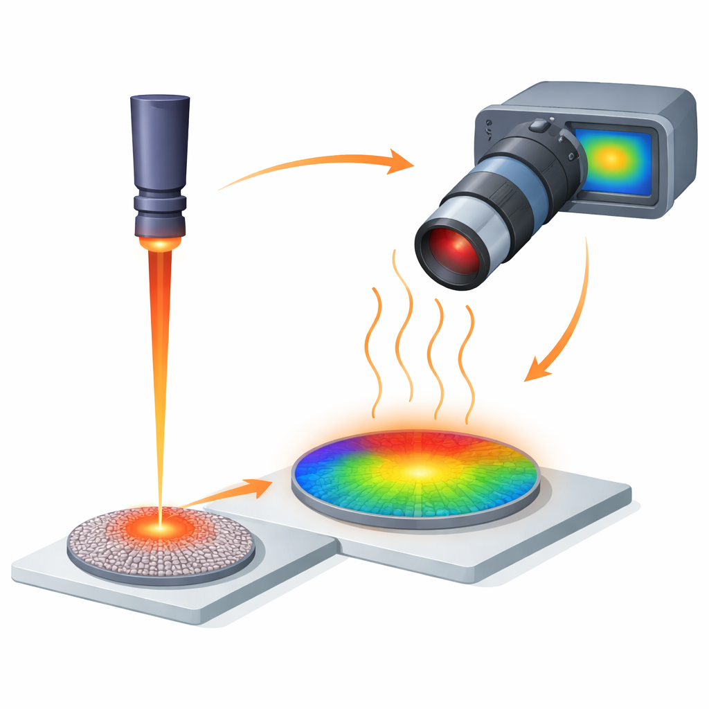
Why Tiny Hotspots Matter
Laser sintering works by focusing a beam onto a bed or pellet of powder so that the particles heat, fuse, and densify into a solid. Especially for semiconductor and oxide nanoparticles such as titanium dioxide, the resulting grain size, pores, and even cracks are governed by the exact temperature history over just a few milliseconds and a few micrometers. Too cool, and the material remains porous; too hot or too long, and it may crack or even ablate. Conventional infrared cameras often lack either the speed or the spatial detail to capture what happens in these tiny hotspots, and thermocouples cannot be placed directly in the active region. The authors therefore turned to near‑infrared light and a high‑speed camera to track temperature without touching the sample.
Turning Glow into Temperature Maps
Any hot object glows in the infrared, and at the high temperatures relevant for laser sintering, a significant part of that glow falls in the near‑infrared range, just beyond visible red. The team modified a commercially available high‑speed camera based on a silicon sensor, removed its built‑in filter, and equipped it with a microscope objective optimized for near‑infrared light. A long‑pass filter blocks visible and ultraviolet light—including the laser itself and any fluorescence—so that the camera only records thermal emission from the heated material. To translate brightness into actual temperature, they carefully calibrated the system using a titanium dioxide pellet heated on a ceramic plate, with the temperature tracked by a thermocouple and a pyrometer. By fitting a standard radiometric equation to these data, they obtained a conversion curve that turns each pixel’s intensity into a temperature, with an accuracy suitable for temperatures between about 600 °C and 900 °C at more than a thousand frames per second.
Zooming in on Fast, Small Hotspots
The microscope optics provide a spatial resolution better than 10 micrometers—fine enough to resolve the approximately 9‑micrometer laser spot on the pellet. Tests with a calibrated microscopic ruler showed that features spaced just 10 micrometers apart could be clearly distinguished, even though the camera views the sample at a 45‑degree angle. At the same time, the camera can record more than a thousand full‑frame images per second, and, with a reduced field of view, nearly sixteen thousand images per second. This combination allowed the researchers to watch the temperature of the hotspot evolve over time as they varied both laser power and pulse duration during resonant ultraviolet laser sintering of titanium dioxide nanoparticles. 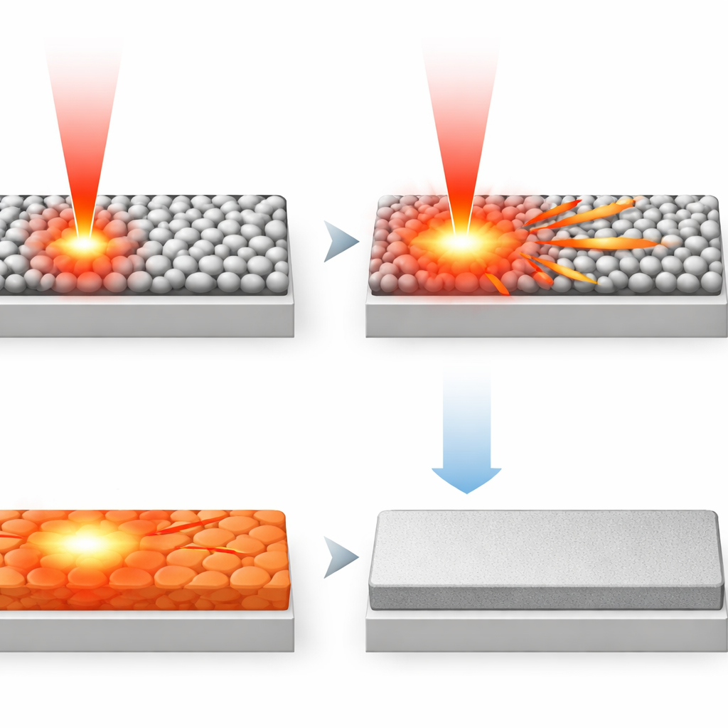
How Heat Shapes the Final Material
With the calibrated system, the team measured how the hotspot temperature responds to laser pulses of different powers and lengths. They found a very rapid temperature rise within the first millisecond of exposure, followed by a slight drop to a plateau that lasts for the rest of the pulse, and then a similarly rapid cooling once the laser is switched off. By adjusting the laser power, they could raise or lower the plateau temperature; by changing the pulse length, they could control how long the material stayed hot. In high‑power experiments, estimated heating and cooling rates reached millions of degrees per second. Scanning electron microscope images of the sintered spots revealed that these temperature‑time profiles directly correlate with the microstructure: moderate powers produced nearly fully dense regions, while higher powers introduced pores, ripples, and eventually cracks or even signs of material removal. The spatial extent of densification matched the region that had experienced the highest measured temperatures.
A New Window into Fast Manufacturing
In everyday terms, the authors have built a high‑speed thermal "microscope" that can watch a tiny patch of material heat up and cool down as a laser fuses nanoparticles into a solid. By linking these detailed temperature movies to the final internal structure, the work shows how manufacturers could tune laser power and timing like knobs to dial in desired properties while avoiding damage. Because the system is compact, based on off‑the‑shelf components, and works at very high temperatures, it could be integrated into a wide range of laser‑based manufacturing setups and even combined with X‑ray instruments. Ultimately, this approach brings us closer to made‑to‑order materials whose structure is shaped with millisecond and micrometer precision.
Citation: Schulte, J., Schroer, M.A. & Winterer, M. Operando high speed near infrared imaging during laser sintering of nanoparticles for time and space resolved temperature measurements. Sci Rep 16, 8158 (2026). https://doi.org/10.1038/s41598-026-37445-7
Keywords: laser sintering, near-infrared imaging, high-speed thermography, nanoparticles, additive manufacturing