Clear Sky Science · en
Flow cytometry helps differentiate between mucous membrane pemphigoid and erosive oral lichen planus
Why painful mouth sores matter
Many people develop long‑lasting sore or peeling gums that make eating and brushing uncomfortable. Two important causes are mucous membrane pemphigoid (MMP) and erosive oral lichen planus (eOLP). They can look remarkably similar in the dentist’s chair, yet they behave differently, need different treatments and carry different long‑term risks. This study explores whether a common laboratory technique, flow cytometry, can read the "fingerprints" of the immune system in a simple blood sample to tell these look‑alike diseases apart.
Two diseases that mimic each other
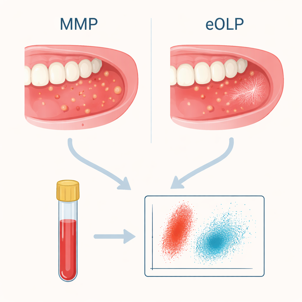
MMP is an autoimmune blistering condition: the immune system makes antibodies that attack the anchoring structures of moist surfaces such as the mouth, eyes, nose and throat. This can lead to fragile blisters, raw erosions and, over time, scarring that may threaten sight or breathing. eOLP, in contrast, is driven mainly by white blood cells called T cells reacting against triggers in the lining of the mouth. It can also cause red, eroded gums and cheeks, sometimes with delicate white lines, and carries a small risk of turning into oral cancer. Because both conditions can present with similar painful erosions on the gums, even tissue samples and standard antibody tests do not always give a clear answer.
Looking at immune cells in the blood
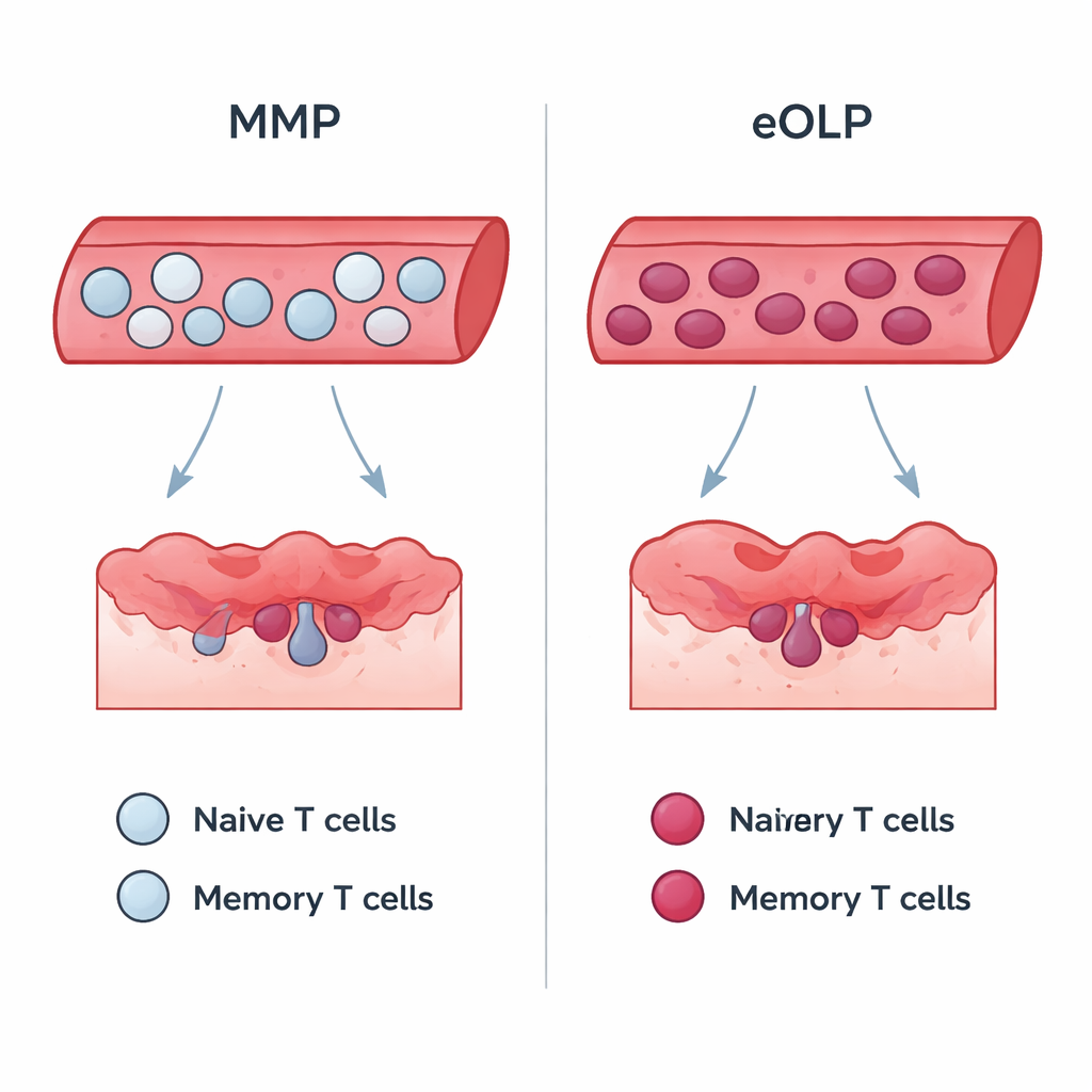
To search for a clearer signal, the researchers enrolled 30 patients: 18 with MMP and 12 with eOLP. All had erosive lesions of the upper and lower gums, and some had additional sites involved. After confirming each diagnosis with accepted clinical, microscopic and antibody criteria, the team drew a blood sample from every participant. Using flow cytometry—a method that passes cells single‑file through a laser to identify their surface markers—they measured many types of B cells (antibody‑producing cells) and T cells (which help or kill other cells), distinguishing "naïve" cells that are new to battle from "memory" cells that have been activated before.
Immune fingerprints that separate the conditions
The B‑cell profiles, which reflect the antibody arm of immunity, were broadly similar in MMP and eOLP. The striking differences lay in certain T‑cell subsets. People with eOLP had higher levels of memory killer T cells and of a helper T‑cell type associated with "type 1" inflammation (often called Th1 cells). Patients with MMP, by contrast, showed more naïve killer T cells and fewer of these memory and Th1 cells. When the authors tested how well these measurements could sort the two diseases, the percentage of memory killer T cells performed best: using a simple cut‑off value correctly classified about 80% of patients, with good balance between sensitivity (catching true eOLP) and specificity (not mislabeling MMP). Th1 cell levels also showed useful, though slightly lower, accuracy.
Clues to disease severity and behavior
Within the MMP group, the researchers also compared patients at higher risk—those with wider involvement such as eyes, throat or genitals—with those whose disease was confined mainly to the mouth. High‑risk patients had more memory helper and killer T cells and more memory B cells, along with fewer naïve cells. This pattern suggests that in more severe MMP, the immune system has been repeatedly activated and is primed to sustain antibody production and tissue damage. Interestingly, these immune patterns did not closely track standard severity scores or blood antibody levels, and similar links were not seen in eOLP, underscoring that the two diseases are driven by different immune mechanics.
What this means for patients and clinicians
For people suffering from chronic sore gums, a clearer diagnosis can guide better care and follow‑up. This work shows that a detailed look at immune cells in a blood sample can highlight differences between MMP and eOLP that routine tests may miss. While the study is small and needs confirmation in larger groups and with healthy controls, it suggests that flow cytometry of T‑cell subsets could become a helpful add‑on test. In everyday terms, the pattern of "seasoned" versus "rookie" immune cells in the bloodstream may act as a bar code that helps doctors distinguish two visually similar but biologically distinct mouth diseases, leading to more tailored treatment and monitoring.
Citation: Liguori, S., Didona, D., Ruoppo, E. et al. Flow cytometry helps differentiate between mucous membrane pemphigoid and erosive oral lichen planus. Sci Rep 16, 6392 (2026). https://doi.org/10.1038/s41598-026-37339-8
Keywords: oral autoimmune diseases, mucous membrane pemphigoid, oral lichen planus, flow cytometry, immune cell profiling