Clear Sky Science · en
Sinus membrane thickness and its correlation with anatomical parameters using cone beam computed tomography: a cross-sectional study
Why the space above your back teeth matters
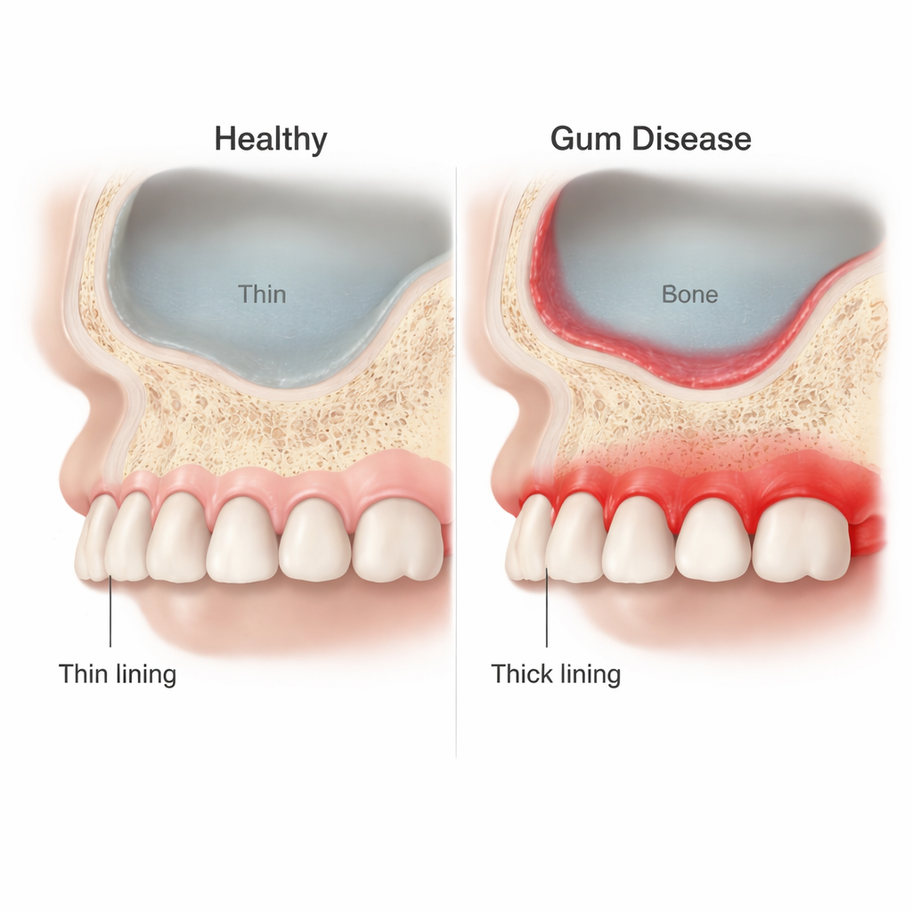
The hollow spaces in our cheeks, known as the maxillary sinuses, sit just above the roots of our upper back teeth. Dentists and surgeons work close to this area when placing dental implants or treating gum disease. If the paper-thin lining of the sinus becomes swollen and thick, surgery can become riskier and sinus problems more likely. This study explores how serious gum disease in the upper jaw may change the thickness and shape of that sinus lining, using detailed three-dimensional scans.
The hidden air pocket above your molars
The maxillary sinus is a pyramid-shaped air cavity in the upper jaw, bordered by the eye socket above and the roots of the back teeth below. Its inside is covered by a soft, moist lining normally less than a millimeter thick. Earlier work has hinted that dental infections and long-standing gum disease can irritate this lining, making it thicker. That matters because a thicker or inflamed lining is more likely to tear during sinus-lift and implant surgeries, and it may block normal drainage, raising the chance of sinusitis. Yet, until now, there have been few studies that carefully measured this lining at several points and compared healthy people with those having advanced gum disease.
Scanning healthy and diseased gums in 3D
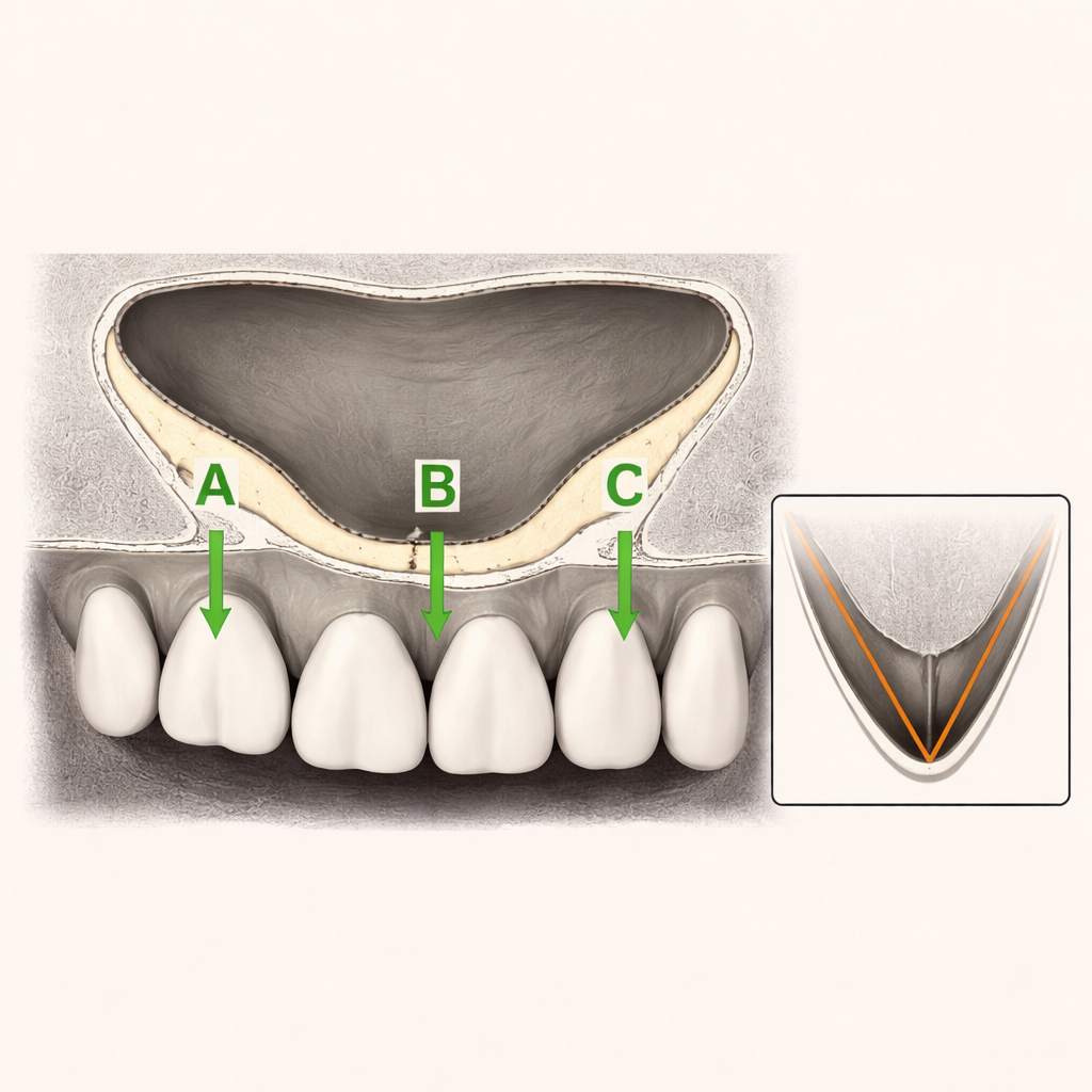
To investigate, the researchers used cone beam computed tomography (CBCT), a type of 3D X-ray widely used in modern dentistry. They recruited 80 generally healthy adults who were already being scanned for dental treatment. Half had healthy gums, while the other half had Stage III periodontitis, a severe form of gum disease that causes deep pockets and bone loss around teeth. On each scan, they measured the thickness of the sinus membrane at three standard points along the sinus floor—front (Point A), middle (Point B), and back (Point C)—on both the right and left sides. They also recorded the overall width of the sinus and the angle formed by its inner walls, features that together describe the sinus’s shape and size.
Thicker lining and altered shape with gum disease
The findings were clear: people with advanced gum disease had a significantly thicker sinus lining at every measured point. For example, at one front point on the right side, the average thickness was about 2.2 millimeters in healthy participants but 3.6 millimeters in those with periodontitis. The single thickest spot anywhere along the sinus floor was also greater in the diseased group. In contrast, the overall width of the sinus cavity was similar in both groups, suggesting that gum disease mainly affects the lining rather than the size of the air space. The angles between the sinus walls were smaller in patients with periodontitis at several points, indicating a subtle change in the inner shape of the sinus that could influence how surgeons approach the area.
Sex, age, and other influences
Across both healthy and diseased groups, men tended to have a thicker sinus lining than women, echoing trends seen in other studies. Some statistical tests also showed links between age, membrane thickness, and sinus angles at specific points, but these relationships were not consistent across all measurements. The authors note that they carefully excluded people with known sinus disease, smoking habits, or allergies that might independently thicken the sinus lining. Even so, they acknowledge that unrecognized sinus problems or other hidden factors could still play a role, and that larger studies using more advanced statistics are needed to separate these effects.
What this means for dental treatment
For patients, the study’s message is straightforward: serious gum disease near the upper back teeth does not just damage the gums and supporting bone; it also tends to thicken and slightly reshape the sinus lining above. This thicker, inflamed tissue is more prone to tearing during sinus-lift and implant procedures and may interfere with normal sinus drainage. The practical takeaway is that careful 3D imaging and thorough gum treatment should come before sinus surgery or implant placement in this region. By calming the gum infection and understanding the exact anatomy of the sinus, dentists and surgeons can plan safer, more predictable treatments with fewer complications.
Citation: Kolte, A.P., Kolte, R.A., Sonare, S.Y. et al. Sinus membrane thickness and its correlation with anatomical parameters using cone beam computed tomography: a cross-sectional study. Sci Rep 16, 6646 (2026). https://doi.org/10.1038/s41598-026-37222-6
Keywords: maxillary sinus, gum disease, dental implants, CBCT imaging, sinus membrane thickness