Clear Sky Science · en
Isolation and proteomic analysis of intracellular vesicles from the potato late blight pathogen Phytophthora infestans
Why tiny bubbles in a blight fungus matter for our food
Potato late blight, caused by the microbe Phytophthora infestans, is the same kind of disease that fueled the Irish potato famine and still destroys crops worth billions of dollars each year. This study looks inside that pathogen at the microscopic “bubbles” it uses to move attack proteins around before they are released into plant tissue. By understanding how these bubbles form, what they carry, and how they travel, researchers hope to find new ways to block infection and protect one of the world’s most important food crops.
The crop killer and its molecular toolkit
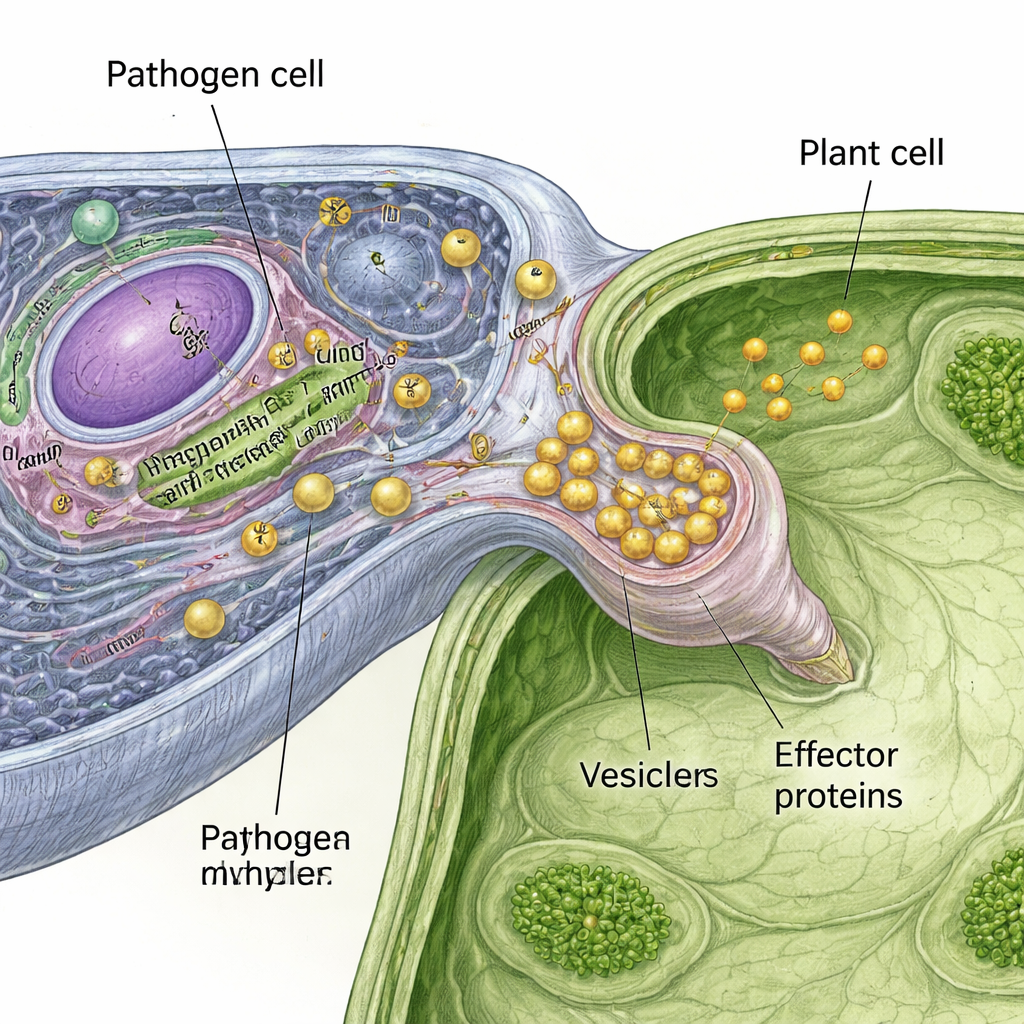
P. infestans is not a true fungus, but it behaves like one, spreading through leaves and stems using thread-like filaments. During infection, it builds special feeding structures, called haustoria, that press into plant cells without breaking them open. At this intimate contact point, the pathogen releases a cocktail of proteins and other molecules that help it slip past plant defenses, digest cell walls and steal nutrients. Many of these proteins are known as effectors. Some work outside plant cells to weaken barriers, while others enter the plant cell interior and rewire its defenses. Although scientists have cataloged many effectors, they have known surprisingly little about how these molecules are packaged and transported inside the pathogen before they are secreted.
Tagging the pathogen’s secret cargo
To watch these pathways in action, the authors engineered P. infestans to make two different effector proteins fused to bright fluorescent tags. One effector represents the well-studied “RXLR” class that enters plant cells, and the other is a pectin-degrading enzyme that acts outside plant cells. Under the microscope, both tagged proteins appeared in tiny bright spots within the pathogen and accumulated at the haustoria during infection of tobacco leaves, suggesting they travel in small membrane-bound bubbles, or vesicles. This gave the team a living marker for secret cargo they could then chase with biochemical methods.
Separating the bubbles without breaking them
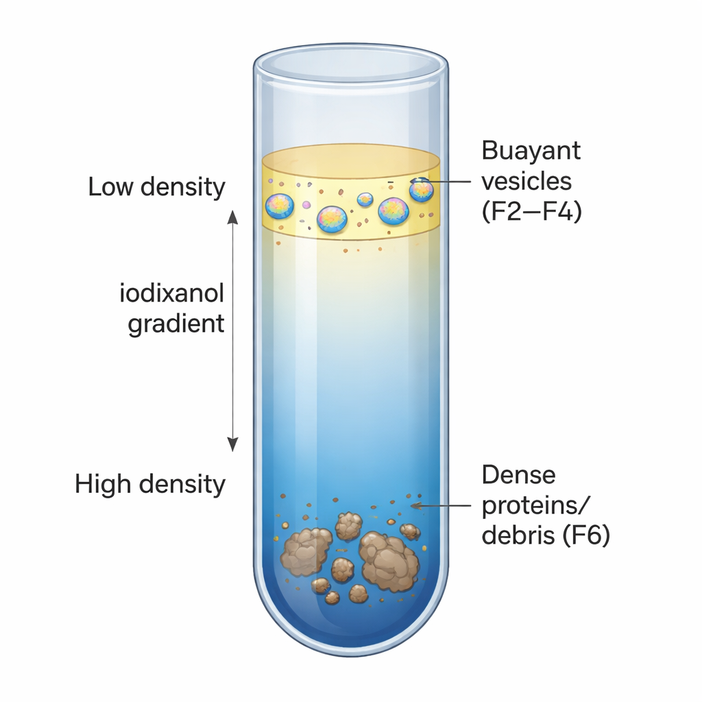
The researchers next developed a careful centrifugation method to fish vesicles out of ground-up pathogen tissue while keeping them intact. They first spun the extract to remove large debris, then floated the remaining material on top of a dense cushion made from iodixanol, a sugar-like compound. A second, long spin through a layered iodixanol gradient allowed structures to settle at the point that matched their natural density. Under these conditions, vesicles collected in lighter “buoyant” layers, while heavier protein clumps and cell fragments sank deeper. Electron microscopy confirmed that the buoyant layers were rich in vesicles, whereas a denser layer, used as a control, contained almost none. When the sample was pre-treated with a detergent that dissolves membranes, the vesicles disappeared and the tagged effectors no longer floated, reinforcing that the method was truly capturing intact bubbles.
What the vesicles are carrying
Using advanced mass spectrometry, the team cataloged more than 6,600 pathogen proteins across the gradient layers and compared those enriched in the buoyant, vesicle-rich fractions with those in the dense control fraction. The vesicle fractions were packed with membrane proteins and secretory proteins bearing signal peptides—molecular zip codes that route cargo toward secretion. They also contained many RXLR effectors, cell-wall-degrading enzymes, and previously reported proteins that mark extracellular vesicles. In contrast, the dense fraction was dominated by housekeeping components such as ribosomal proteins and enzymes for gene expression, consistent with leaked cell contents rather than transport bubbles. Further comparisons between lighter and slightly heavier vesicle fractions showed each contained distinct sets of proteins linked to different cellular locations, hinting at multiple specialized vesicle types that may shuttle effectors along specific routes.
Turning basic insight into better blight control
For non-specialists, the key message is that the authors have built a reliable way to isolate and profile the microscopic bubbles that move attack proteins inside P. infestans. Their protein catalogue reveals both the membranes that form these vesicles and the cargo they carry, including many molecules directly involved in disease. This framework will allow future work to trace how effectors are sorted, packaged and dispatched from pathogen to plant. In the long run, targeting the machinery that builds or steers these bubbles could offer new strategies to stop late blight—not by killing the pathogen outright, but by cutting the supply lines it needs to invade and damage potato crops.
Citation: Pham, J., Whisson, S.C., Hurst, C.H. et al. Isolation and proteomic analysis of intracellular vesicles from the potato late blight pathogen Phytophthora infestans. Sci Rep 16, 6185 (2026). https://doi.org/10.1038/s41598-026-37161-2
Keywords: potato late blight, Phytophthora infestans, effector proteins, intracellular vesicles, plant disease control