Clear Sky Science · en
Breast imaging with ultra-low field MRI
A New Way to Look Inside the Breast
Most women are told to get regular breast cancer screenings, yet many skip them because of discomfort, cost, or limited access to high-tech scanners. This study explores a very different kind of breast scan: an "ultra-low field" MRI system that is quieter, simpler, and potentially far cheaper than hospital MRI, while avoiding the X‑ray radiation used in mammograms. The researchers set out to see a basic but crucial question—can such a gentle, low-power scanner still show the key structures inside the breast well enough to be useful one day for screening?
Why Current Screening Tools Aren’t Enough
Today, mammography is the workhorse of breast cancer screening worldwide. It is widely available and relatively inexpensive, but it has real drawbacks. It uses ionizing radiation, which many women worry about over repeated exams, and it requires the breast to be squeezed between plates, which can be painful. Mammograms can also miss cancers, especially in women with dense breast tissue, and they generate a sizable number of false alarms that lead to stress and extra tests. MRI scans can see soft tissues much more clearly and do not use radiation, but standard MRI machines are huge, expensive, and scarce, and typical breast MRI exams require an injection into a vein. As a result, MRI is reserved for women at particularly high risk, leaving most of the global population reliant on mammography alone.
What Ultra-Low Field MRI Tries to Do Differently
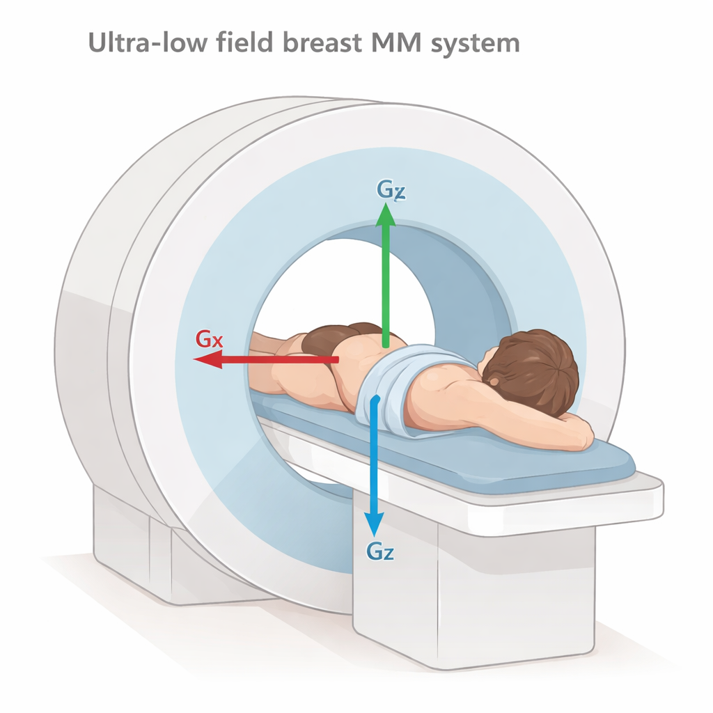
The team behind this study built a custom MRI scanner that uses a magnetic field roughly 200 times weaker than a standard hospital MRI. Lowering the field strength lets them replace the massive superconducting magnet with a far simpler, less demanding setup. They also created a snug, cone-shaped coil that surrounds just one breast at a time, improving the signal they can capture. In their exam, a woman lies face down on a table with one breast resting naturally inside this cone—no compression, no needles, and no contrast dye. The scanner then collects a three-dimensional series of images in about 20 to 45 minutes, depending on how detailed the pictures need to be.
What the Early Images Reveal
To test the idea, the researchers scanned 11 healthy women and three patients who had either previous breast cancer or a known benign cyst. Three independent breast radiologists examined the ultra-low field images and rated whether they could see essential features: the overall shape of the breast, the internal fibroglandular tissue (the denser, milk-producing parts), the fatty areas, the nipple–areolar region, and the chest wall muscle behind the breast. Across the healthy volunteers, the readers consistently identified the breast outline and separated fatty from fibroglandular tissue patterns, even at modest resolution. In a subset of women who also had recent mammograms, the new MRI method showed tissue patterns that matched what the X‑ray images revealed, suggesting it is capturing the same broad anatomy in a very different way.
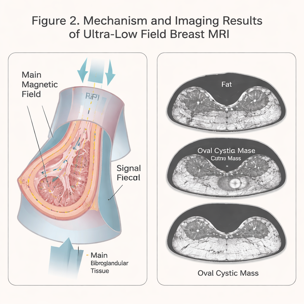
Seeing Scars and Cysts Without Metal Artifacts
The three patient cases hint at what ultra-low field MRI might add beyond basic anatomy. Two women had undergone lumpectomy for breast cancer, leaving surgical clips and scar tissue behind. On standard high‑field MRI, those metal clips cause dark, blooming artifacts that can hide nearby tissue. At ultra-low field, the same clips produced almost no distortion, yet the scar lines were still visible, allowing the radiologist to see post-surgical changes without losing surrounding detail. In the third patient, who had a large, fluid-filled cyst already confirmed by ultrasound, the cyst appeared clearly on several ultra-low field slices, and its measured size closely matched the ultrasound findings. That suggests that even with lower resolution, sizable fluid-filled lumps may be detectable and measurable.
Limits Today and Hopes for Tomorrow
This work is an early technical demonstration, not a ready-made screening test. The images are still coarser than what is required to reliably spot small cancers, and only one breast can be scanned at a time, with exam times that are longer than ideal. Some structures, such as the chest wall in women with larger breasts and the nipple area in certain cases, were not always seen clearly, pointing to the need for better coil designs and faster imaging methods. The study also included very few patients and no untreated cancers, so it cannot yet tell us how well this approach would actually detect disease. Still, the results show that a simple, ultra-low field system can capture meaningful breast anatomy, surgical changes, and at least one benign mass without radiation, compression, or injections. With further engineering and clinical testing, such scanners could one day offer a more comfortable, accessible option that brings advanced breast imaging closer to everyday clinics and resource-limited settings.
Citation: Shen, S., Koonjoo, N., Longarino, F.K. et al. Breast imaging with ultra-low field MRI. Sci Rep 16, 4518 (2026). https://doi.org/10.1038/s41598-026-37130-9
Keywords: breast cancer screening, ultra-low field MRI, breast imaging, mammography alternatives, medical imaging technology