Clear Sky Science · en
¹H-NMR serum metabolomic profiling from clinical routine identifies signatures of progressive melanoma metastasis
Why Blood Chemistry Matters in Skin Cancer
For people living with melanoma, one of the deadliest forms of skin cancer, the biggest worry is whether the disease has quietly spread to other parts of the body. Today, doctors rely mainly on scans and a few blood tests to spot this spread, but these tools often detect trouble only once tumors are large or numerous. This study explores whether the chemical fingerprints in a simple blood sample can reveal, much earlier, when melanoma has become actively metastatic—and does so using samples collected during everyday hospital care, not in a carefully controlled lab setting.
Looking for Hidden Signals in the Blood
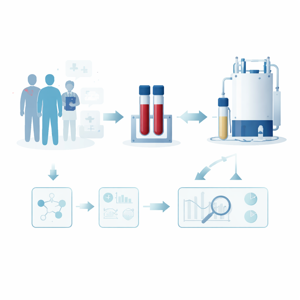
The researchers focused on molecules called metabolites, the small chemical building blocks and fuels that constantly flow through our bodies. Cancer cells are known to rewire how they use energy and nutrients, and this metabolic reshaping can spill over into the bloodstream. The team collected 1,698 serum samples from 963 melanoma patients treated at a German university hospital. Using a technique called proton nuclear magnetic resonance (¹H-NMR) spectroscopy, they measured dozens of soluble metabolites in each sample and then asked a simple question: do patients with currently active metastases show a distinct chemical pattern in their blood compared with those whose disease is not actively spreading?
Turning Complex Data into a Risk Score
To make sense of this rich chemical information, the scientists applied advanced statistical and machine-learning tools. They split patients into two groups: one to build their prediction models and another, kept strictly separate, to test how well those models worked in practice. Two different approaches were used to pick out the most informative metabolites and to combine them into a single score that estimates whether a patient has active metastatic disease. When tested on the independent group, these models could distinguish active metastasis from non-active disease better than chance, but not with perfect accuracy. Their performance landed in a moderate range, suggesting they can detect a real biological signal but are not yet reliable enough to stand alone in clinical decision-making.
What Changes in the Blood Reveal
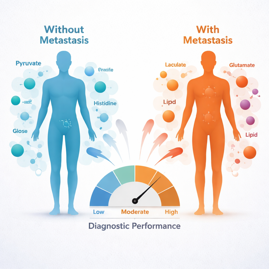
Despite only moderate predictive power, the patterns themselves were striking. Patients with active metastases tended to have higher blood levels of pyruvate, glucose, glutamate, acetoacetate, and the amino acid phenylalanine, and lower levels of histidine and citrate. In everyday terms, this points to a broad reshaping of how energy is produced and how amino acids are used in the body. Elevated pyruvate and glucose hint at cancer-driven changes in sugar use and energy generation, while shifts in citrate and acetoacetate point to altered activity of key energy cycles and fat metabolism. Reduced histidine and changes in other amino acids suggest that tumors may be drawing heavily on specific building blocks from the bloodstream to fuel growth and spread.
Special Treatment and Tumor Types Leave Their Mark
The team also explored whether specific clinical situations leave their own metabolic footprints in blood. Among patients receiving modern immunotherapies, such as immune checkpoint inhibitors, those on these treatments showed different levels of certain metabolites, including citrate, compared with patients on other systemic drugs. In people whose melanoma had already spread, subtle blood chemistry differences were seen between tumors that had reached the brain and those confined to other organs, and between tumors with or without a common mutation in the BRAF gene. These subgroup findings were modest and exploratory, but they suggest that both the cancer’s genetics and the type of treatment may shape the metabolic signatures seen in blood.
What This Means for Patients and Future Care
For someone facing melanoma, the main message is that a routine blood draw may one day do more than check general health—it could help reveal whether the cancer is actively spreading and how the disease and its treatment are reshaping the body’s chemistry. This study shows that such information is indeed present in the blood and can be detected even under real-world conditions, but the current tests are not yet accurate enough to replace scans or existing biomarkers. Instead, the authors see these metabolic patterns as a promising piece of a larger puzzle. Combined with imaging, genetic tests, and other blood markers, metabolite profiles like those centered on pyruvate and histidine could form part of more powerful, multi-tool panels that better track melanoma and guide treatment choices.
Citation: Gellrich, F.F., Hufnagel, C., Funk, A.M. et al. ¹H-NMR serum metabolomic profiling from clinical routine identifies signatures of progressive melanoma metastasis. Sci Rep 16, 5263 (2026). https://doi.org/10.1038/s41598-026-37118-5
Keywords: melanoma, metastasis, serum metabolomics, cancer biomarkers, NMR spectroscopy