Clear Sky Science · en
Lung ultrasound B-line quantification in CTD-ILD: a cross-sectional single-center observational study
Scanning the Lungs Without Radiation
People living with autoimmune diseases often worry about their lungs. These conditions can quietly scar lung tissue over time, making it harder to breathe, yet the main test to see this damage—high‑resolution CT scanning—uses radiation and can be costly. This study asks a simple, patient‑friendly question: can a quick ultrasound scan over the chest, much like the one used in pregnancy, give doctors a reliable picture of how badly the lungs are affected?
A Gentle Look Inside Troubled Lungs
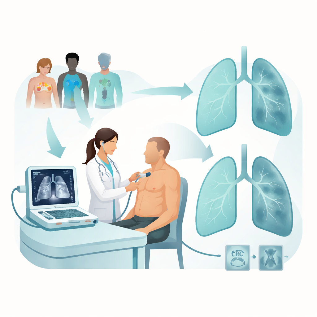
Connective tissue diseases—such as rheumatoid arthritis, systemic sclerosis, lupus, and Sjögren’s syndrome—can inflame and scar the delicate air sacs of the lungs, a condition known as interstitial lung disease. Patients may notice cough, breathlessness, and fatigue, but by the time symptoms are obvious, lung damage can already be advanced. CT scans reveal this damage in detail, yet repeated scans are not ideal for long‑term monitoring. Lung ultrasound, by contrast, is portable, safe, and repeatable at the bedside. When the normally air‑filled tissue beneath the lung surface becomes water‑logged or scarred, it creates bright vertical streaks on ultrasound images called B‑lines. Counting these streaks may offer a simple way to estimate how much of the lung is affected.
How the Study Tested Ultrasound Scoring
The researchers enrolled 117 adults with both a connective tissue disease and confirmed interstitial lung disease. Within a single week, each person underwent lung ultrasound, a CT scan, lung function testing, and blood work. The chest was divided into 12 regions, and each region was scored from 0 to 3 depending on how many B‑lines or solid areas appeared, giving an overall ultrasound score from 0 to 36. Higher scores meant more abnormal lung tissue near the surface. CT scans were graded with an established scale that rates how much scarring and inflammation is seen, and patients were grouped as having mild, moderate, or severe lung involvement. Standard breathing tests measured how much air patients could blow out and how well oxygen could pass from the lungs into the blood.
What Ultrasound Revealed About Lung Damage
People with higher ultrasound scores tended to have worse CT findings and poorer breathing test results. In particular, higher scores were linked to lower amounts of air exhaled in one second and more extensive damage seen on CT images. When the team explored a simple cut‑off point on the ultrasound scale, patients above that threshold generally had more severe lung changes on CT, weaker breathing numbers, and certain signs of immune system activation in their blood. The link between ultrasound and CT was especially strong in patients with Sjögren’s syndrome, suggesting that ultrasound may be particularly informative in this subgroup. However, in other autoimmune diseases, such as systemic sclerosis or inflammatory muscle disease, the relationship between ultrasound and CT was weaker or less consistent.
How Well Can Ultrasound Sort Mild From Severe Disease?
The investigators then asked how well ultrasound alone could sort patients into mild, moderate, or severe lung disease. Ultrasound scores did a reasonable job at flagging those with clearly severe damage, but they struggled to cleanly separate mild from moderate cases. When the ultrasound score was combined with a key lung function measure that reflects how well oxygen crosses into the blood, the ability to distinguish mild from severe disease improved noticeably. This finding underscores that a single bedside test is rarely enough; instead, combining structural clues from imaging with functional measures of breathing provides a fuller picture of lung health.
What This Means for Patients and Doctors
For people with connective tissue diseases, this work supports lung ultrasound as a practical, radiation‑free tool to monitor lung involvement over time. A rising B‑line score can alert clinicians that scarring or inflammation near the lung surface is worsening, prompting closer follow‑up or treatment changes without immediately resorting to another CT scan. Still, ultrasound cannot fully replace CT or breathing tests, especially when doctors need to tell moderate from severe disease or to map deeper lung regions. The study suggests that ultrasound works best as part of a toolkit, alongside CT and lung function testing, to guide decisions and reduce unnecessary radiation while keeping a close eye on a vital organ.
Citation: Du, M., Wang, J., Lai, P. et al. Lung ultrasound B-line quantification in CTD-ILD: a cross-sectional single-center observational study. Sci Rep 16, 6099 (2026). https://doi.org/10.1038/s41598-026-36874-8
Keywords: lung ultrasound, interstitial lung disease, connective tissue disease, B-lines, noninvasive imaging