Clear Sky Science · en
High-fidelity and efficient particle microscopy via regional focus search and adaptive focus stacking
Why Sharper Tiny Particles Matter
From drug-delivery nanoparticles to industrial powders, many modern technologies depend on tiny particles whose size and shape must be measured with great precision. Yet even the best microscopes have a built-in weakness: they can only keep a thin slice of depth sharply in focus at once. When particles sit at different heights in a droplet or on a slide, some look crisp while others blur, and that blur can seriously distort measurements. This article presents a practical way to turn a stack of imperfect microscope images into one clear, all-in-focus picture that captures particle size and shape much more accurately.
Blurry Pictures, Misleading Measurements
Under a microscope, micro- and nanoparticles rarely sit neatly on a single flat plane. Instead, they are scattered at slightly different depths. Because a lens can only sharply image a narrow layer at once, parts of the picture are in focus while others are not. When image-analysis software tries to measure particle size and roundness from such mixed-quality images, it tends to overestimate sizes and underestimate how circular the particles are. These errors are not just cosmetic; they can influence how a drug is released in the body, how a catalyst behaves, or how a material wears over time. The authors focus on standard polystyrene beads a few micrometers across, but the problem and solution are broadly relevant to many fields.
Building a Clearer Composite Image
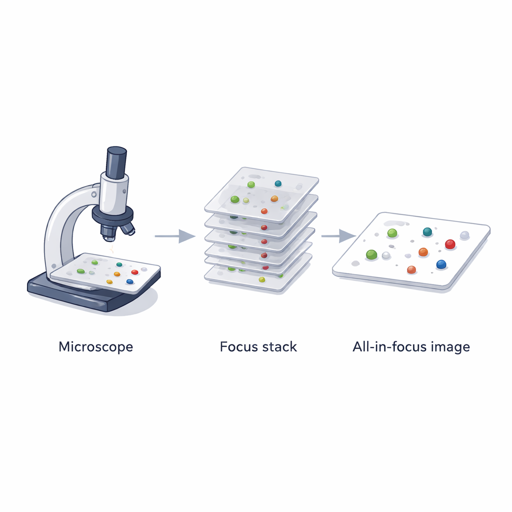
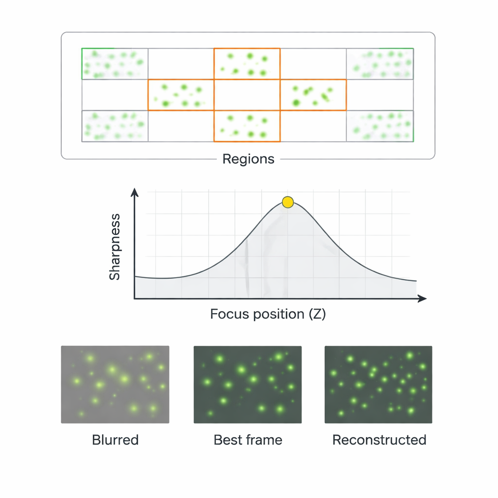
To overcome this depth-of-field limitation, the researchers combine two ideas: a smarter way to find the best focus, and an image-fusion step called focus stacking. First, they evaluate how sharp an image is using a simple statistical measure of gray-level variation (the variance), which reliably tracks how crisp particle edges appear. Next, they scan the sample in the up–down direction, collecting a stack of images at different focus positions. Instead of treating every pixel equally, they automatically locate regions that likely contain particles and concentrate their analysis there, ignoring background areas that only add noise and computation time. For each of these regions, the method searches along the focus axis to find the position where the particles look sharpest, then stitches together those best-focused patches into a single all-in-focus image.
Smarter Focus and Better Size Standards
The team also refines how particle size is defined so that measurements remain stable even when microscope settings like brightness or contrast change. They compare several common size metrics and find that two are especially robust: one based on the shortest distance across a particle, and one based on the area of its outline. Their rule of thumb is intuitive: when a particle is nearly round, they use the shortest axis as its size; when it is irregular, they switch to the area-based diameter. This adaptive standard better reflects the true geometry of both isolated and clumped particles. In parallel, they speed up focusing by combining a quick, coarse search along the full depth range with a slower, fine-grained search only inside particle regions, cutting focus-search time by more than a factor of four while preserving accuracy.
Sharper Images, Smaller Errors
The researchers put their approach to the test on mixtures of polystyrene beads with known diameters. They captured dozens of images at different focus levels and compared three cases: a single frame that looked best by eye, a poorly focused frame, and their reconstructed all-in-focus image. When they measured particle sizes from these images, the reconstructed version yielded errors of only about 1–2% on average, far below the roughly 5–14% errors from single frames. For clumped particles, where outlines are harder to see, the method also cut shape errors by more than two-thirds. Importantly, the technique worked not only for one bead size but also for mixtures of different sizes, allowing the team to resolve distinct size peaks in the distribution that would otherwise blur together.
What This Means for Real-World Labs
In practical terms, this work shows that laboratories can dramatically improve the reliability of particle measurements without buying exotic new microscopes or training large deep-learning models. By using a simple sharpness measure, a targeted focus search, and a careful definition of particle size, the method turns a stack of ordinary microscope images into a high-fidelity, all-in-focus view. For scientists and engineers, this means more trustworthy particle size and shape data, clearer separation between different size groups in mixtures, and better links between microscopic structure and real-world performance of materials, drugs, and devices.
Citation: Xu, C., Tao, Y., Guo, X. et al. High-fidelity and efficient particle microscopy via regional focus search and adaptive focus stacking. Sci Rep 16, 5755 (2026). https://doi.org/10.1038/s41598-026-36757-y
Keywords: particle microscopy, focus stacking, image sharpness, particle size analysis, micro-nano particles