Clear Sky Science · en
Dysfunctional connectivity within hippocampal and entorhinal networks underlies early-life iron deficiency induced social recognition deficits, a preliminary study
Why a lack of iron in early life matters for the brain
Iron deficiency is usually talked about in terms of tiredness and anemia, but for babies and young children it can quietly reshape the developing brain. This study used young rats to ask a specific question: does a shortage of iron early in life disturb the brain circuits that allow animals to recognize and respond to other individuals? By combining behavioral tests with advanced brain imaging, the researchers traced how low iron alters communication between key memory and social areas deep in the brain.
Getting a closer look with animal models
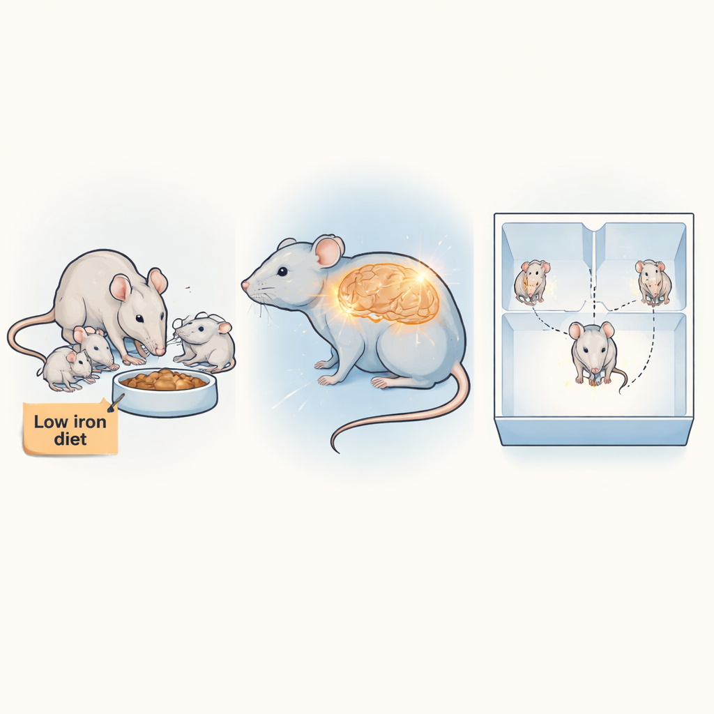
Human studies have linked early-life iron deficiency with lasting problems in thinking, mood, and social behavior, but they face major obstacles: scans are done late, cannot be repeated often, and results are tangled with differences in family life, income, and genetics. To sidestep these issues, the team raised rats on either a normal diet or a very low-iron diet from before birth until five weeks of age, roughly comparable to preteen years in humans. This controlled setup let them ask how iron shortage alone alters brain structure and function, while also testing the animals’ social skills and basic movement.
Social missteps without motor problems
To probe social behavior, the researchers used a three-chamber box. In one phase, a rat could choose between an unfamiliar rat and an empty chamber; in another, it chose between a familiar rat and a new stranger. Both healthy and iron-deficient rats preferred another rat over an empty space, showing normal sociability. The key difference emerged in the second phase: healthy rats spent more time with the new stranger, showing they could recognize and distinguish the familiar animal, while iron-deficient rats failed to show this preference. Their social recognition was blunted, even though tests of balance, grip strength, and walking showed that their movement and coordination were normal.
Brain regions grow larger but talk less
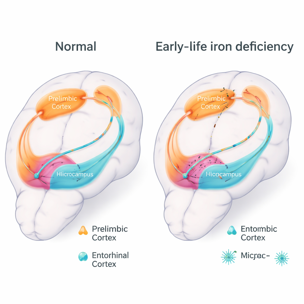
The team then turned to high-field MRI scans. A structural analysis called voxel-based morphometry revealed that two deep brain regions crucial for memory and navigation—the hippocampus and the neighboring entorhinal cortex—were actually enlarged in iron-deficient rats, rather than shrunken. Microscopic staining did not show a loss of nerve cells, but it did reveal more activated microglia, the brain’s immune sentinels, hinting at subtle inflammation or remodeling. Resting-state functional MRI, which tracks how brain regions’ activity rises and falls together, told a different story: connections between the hippocampus and entorhinal cortex were markedly weaker, and their links to a frontal area involved in planning and social behavior, the prelimbic cortex, were also reduced.
Pinpointing weak links in the memory–social network
To move beyond broad regions, the researchers divided the hippocampus and entorhinal cortex into smaller, known subregions and mapped how strongly each pair communicated. This “deep” connectivity analysis showed that the lateral entorhinal cortex—a gateway that feeds detailed information about experiences into the hippocampus—was especially affected. Its connections with several hippocampal subregions, including the dentate gyrus and subiculum, were severely weakened. At the same time, connections within the entorhinal cortex itself were disrupted. Together, these changes suggest that early iron deficiency leaves the main highway for processing and relaying social and memory information partly broken, even though the roadways are still physically present.
What this means for children at risk
In everyday terms, the study suggests that a lack of iron early in life does not simply slow brain growth; it scrambles how key memory and social hubs talk to one another. The affected rats could move normally, but they struggled with a subtle social task that relies on recognizing who is who. Because the same brain regions and networks support social memory in humans, these findings strengthen concerns that early iron deficiency may quietly undermine social and emotional development. They also show that MRI can pick up telltale patterns of altered brain connectivity, hinting at future tools for early detection—and underscoring the importance of preventing iron deficiency in mothers and young children.
Citation: Ding, A., Tan, T., Liu, P. et al. Dysfunctional connectivity within hippocampal and entorhinal networks underlies early-life iron deficiency induced social recognition deficits, a preliminary study. Sci Rep 16, 6474 (2026). https://doi.org/10.1038/s41598-026-36710-z
Keywords: early-life iron deficiency, social recognition, hippocampus, functional connectivity, resting-state fMRI