Clear Sky Science · en
Analysis of the spatial-temporal distribution and functional morphology of telocytes in goat testes
Hidden Helpers in Male Fertility
Deep inside the testes of mammals, fertility depends on a finely tuned neighborhood of cells that support developing sperm. This study uncovers a little-known cell type, called telocytes, in the testes of goats and shows how they change as animals grow from infancy to adulthood. Understanding these cells may eventually help veterinarians and researchers protect male fertility in livestock and, by extension, offer clues for human reproductive health.
A New Cell Type in a Busy Neighborhood
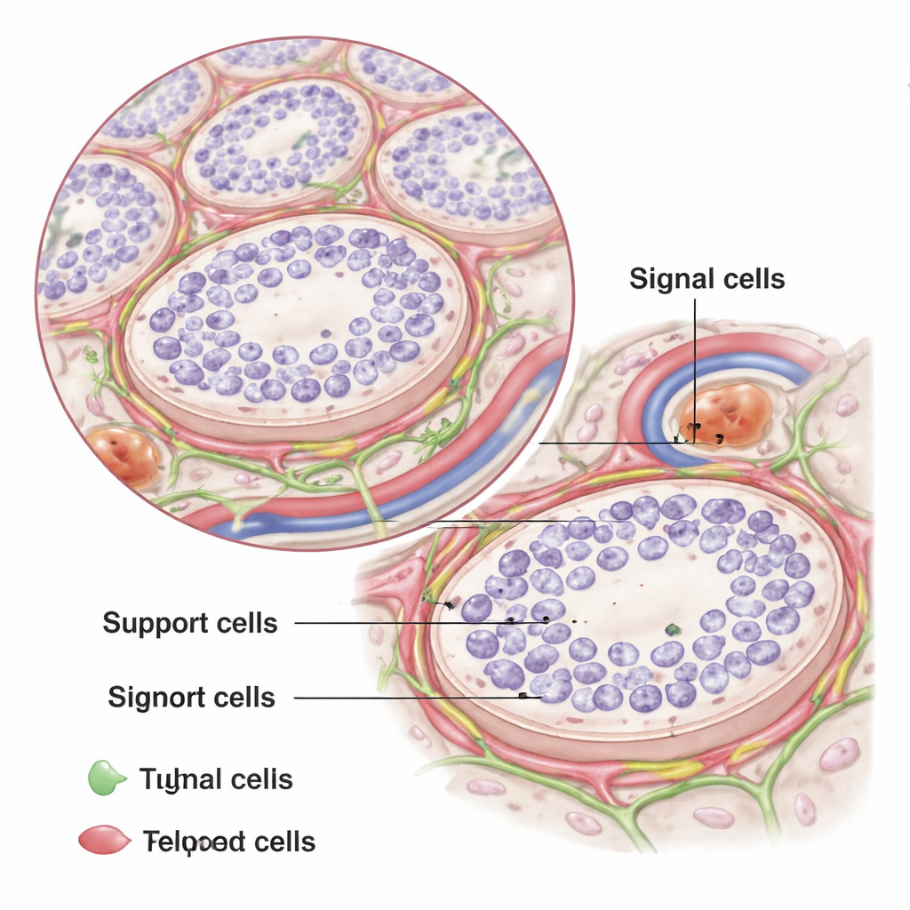
The testes are packed with tiny tubes where sperm develop, surrounded by support cells, blood vessels, and structural tissue. The authors set out to find out whether telocytes – long, slender cells first discovered in other organs – also exist in goat testes. Telocytes have tiny cell bodies and extremely long, thread-like extensions that can stretch tens to hundreds of micrometers, forming a kind of communication and support network. Until now, no one had confirmed their presence in goat testes or described how they are arranged at different ages.
Looking Close: From Electron Beams to Glowing Markers
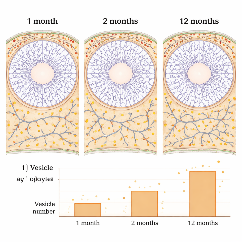
To track down these elusive cells, the team studied testes from goats that were one month, two months, and twelve months old, covering pre-puberty, the onset of sperm production, and full sexual maturity. They used transmission electron microscopy, which shoots electrons through ultra-thin tissue slices to reveal structures at very high resolution, and double immunofluorescence, which tags specific proteins with glowing dyes. Together, these methods allowed the researchers to recognize telocytes both by their shape and by the protein markers on their surface.
How Telocytes Are Built and Where They Sit
Under the electron microscope, telocytes showed their signature look: very small cell bodies and extremely long, thin extensions with a beaded appearance. These extensions, called telopodes, were made of alternating narrow segments and slightly swollen segments that held cell machinery and tiny vesicles. In the youngest goats, telocytes were few and formed only a couple of delicate layers around the sperm-forming tubules. By twelve months, however, they formed four to five layers outside the muscle-like peritubular cells and wove among collagen fibers and blood vessels, creating intricate networks. The telopodes frequently touched other cell types, including supportive muscle-like cells, stromal cells, and hormone-producing Leydig cells, suggesting that telocytes are well placed to sense and relay signals in the testicular tissue.
Signs of Growing Activity with Age
Immunofluorescence experiments helped pin down the identity of these cells. The authors showed that goat testicular telocytes consistently carried the surface protein CD34 and the internal filament protein vimentin, but did not appear to use another common marker, PDGFR-α, in this species. As goats matured, telocytes became longer, their nuclei more elongated, and their projections more numerous, forming denser networks. At the same time, the researchers counted a sharp rise in the number of tiny extracellular vesicles – small, membrane-bound packages – clustered around telopodes in adult goats. These vesicles are known in other systems to ferry signals and molecules between cells, hinting that telocytes in mature testes may communicate more intensely and play a larger role in shaping the local environment.
Why These Cells Matter for Fertility
Because telocytes sit between the contractile muscle-like layer and the wider testicular tissue, and because their networks surround both sperm-forming tubules and nearby blood vessels, the authors propose that these cells help stabilize the spermatogonial stem cell niche – the specialized home where the earliest sperm-forming cells reside. By providing structural support, helping to organize the surrounding matrix, and releasing signaling vesicles, telocytes may help maintain the barrier that protects developing sperm and keep the testicular microenvironment in balance. While this study does not yet prove exactly how telocytes influence fertility, it firmly establishes their presence, their distinct features, and their age-related changes in goat testes, laying the groundwork for future work on how these “signal cells” might become targets for improving or preserving male reproductive health in animals and possibly humans.
Citation: Feng, J., Dai, C., Wang, Q. et al. Analysis of the spatial-temporal distribution and functional morphology of telocytes in goat testes. Sci Rep 16, 5790 (2026). https://doi.org/10.1038/s41598-026-36639-3
Keywords: male fertility, telocytes, goat testis, stem cell niche, testicular microenvironment