Clear Sky Science · en
Visual classification of allergenic pollen in iteratively reconstructed lens-less DIHM images
Why tiny grains of pollen matter
Every spring, countless dogs – and their owners – struggle with itchy skin and allergies triggered by airborne pollen. Pinpointing exactly which plant species are to blame usually requires bulky, expensive microscopes and trained specialists. This study asks a simple but powerful question: can a cheap, lens-free imaging gadget, paired with smart computer processing, give experts images good enough to recognize troublesome pollen grains by eye, without a traditional microscope?
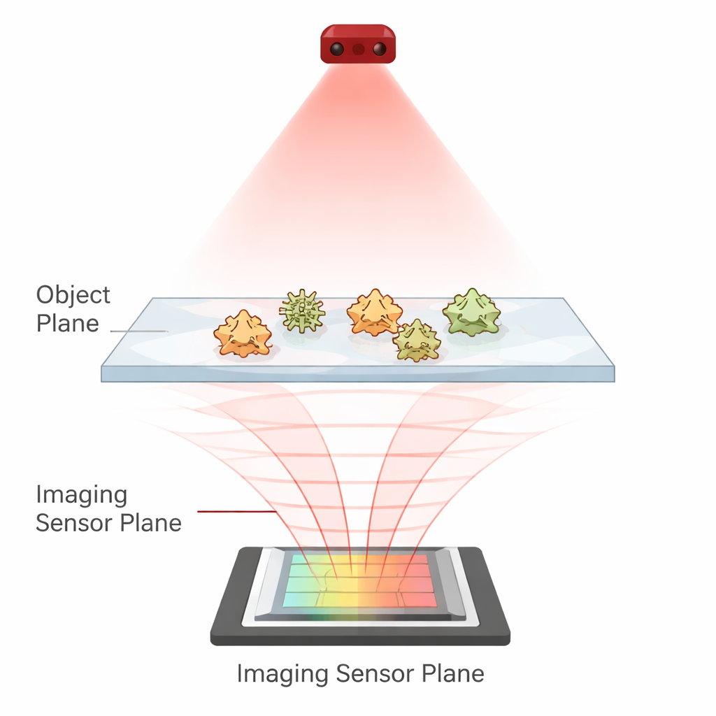
A microscope without lenses
Instead of using glass lenses to magnify tiny objects, lens-less digital in-line holographic microscopy (DIHM) shines a laser through a sample directly onto a camera chip. The pollen grains slightly disturb the light, creating a delicate interference pattern – a hologram – on the sensor. On its own, this pattern looks nothing like a familiar microscope picture. But with the right mathematics, it can be “refocused” on a computer to reveal the pollen’s shape. The catch is that a simple refocusing step produces a sharp image of each grain surrounded by a fuzzy echo, known as a twin image, which lowers contrast and makes visual evaluation harder.
Cleaning up the picture with iteration
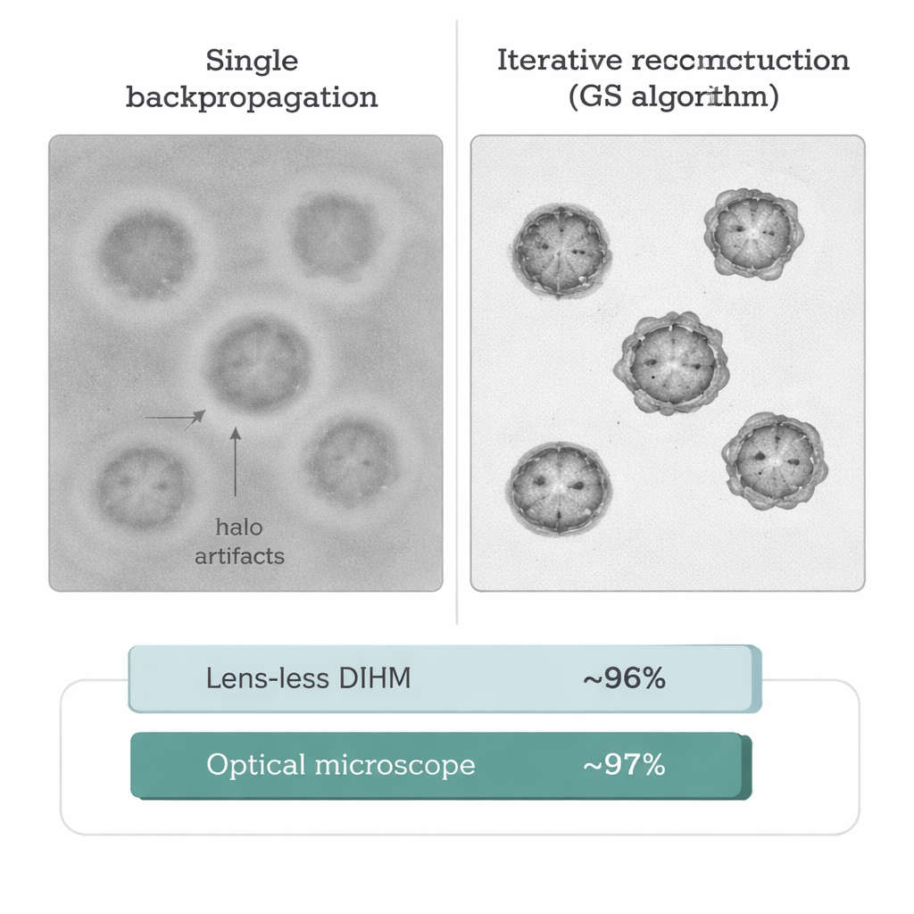
To overcome this problem, the researchers used an iterative computer method called the Gerchberg–Saxton (GS) algorithm. Starting from the recorded hologram, the algorithm repeatedly shuttles the light field back and forth between the camera plane and the pollen plane in a virtual sense, each time enforcing simple physical rules such as “the sample cannot brighten light beyond its original intensity.” After about 200 such cycles, the unwanted twin image is largely removed. The resulting picture looks much more like a standard brightfield microscope image: the background is cleaner, edges of pollen grains are crisp, and key shape features are clearly visible.
Putting human experts to the test
To see whether these cleaned-up, lens-less images are truly useful in practice, two veterinary cytopathologists – doctors who routinely interpret microscopic samples from animals – were asked to classify common allergenic pollen types. The study focused on six species known to trigger skin disease in dogs, including timothy grass, common ragweed, silver birch, common alder, olive tree, and hazel. For each of 60 slides, the same areas were imaged in two ways: once with the lens-less DIHM system and once with a conventional optical microscope. The experts, working under realistic viewing conditions on a standard computer, had to identify which plant each image came from, using only their eyes and a small set of reference pictures.
How well did the lens-free system perform?
The results were striking. With lens-less DIHM images, the overall classification accuracy reached 95.8%; with a conventional optical microscope, it was 96.9%. In practical terms, this difference amounted to only one extra misclassified sample. Agreement between the two experts was also extremely high (Cohen’s kappa of 0.91), showing that both the new and old imaging methods supported consistent judgments. Most mistakes occurred when distinguishing between silver birch, alder, and hazel, whose pollen grains can share similar triangular or polygonal shapes. Four other pollen types, including timothy grass and common ragweed, were almost always recognized correctly, regardless of the imaging method, thanks to their more distinctive outlines and surface features.
What this means for pets and people
For general readers, the key message is that a compact, inexpensive, lens-free device can produce computer-reconstructed images that human experts find nearly as reliable as those from a traditional laboratory microscope. In veterinary clinics and other resource-limited settings, such a system could eventually help identify which pollens individual animals – or even people – are exposed to, without the need for large, delicate optical instruments. While the study was limited to a controlled set of samples and a handful of pollen types, it shows that carefully processed holographic images are clear enough for expert eyes, opening the door to portable allergy monitoring tools that could fit on a benchtop – or even in the field.
Citation: Cugmas, B., Štruc, E., Tamosiunas, M. et al. Visual classification of allergenic pollen in iteratively reconstructed lens-less DIHM images. Sci Rep 16, 6006 (2026). https://doi.org/10.1038/s41598-026-36618-8
Keywords: pollen allergies, lensless microscopy, holographic imaging, veterinary dermatology, allergen monitoring