Clear Sky Science · en
A rabbit model of clinically relevant mucosal injury induced by nasogastric tube intubation
Why a feeding tube can hurt the nose
Feeding through a tube in the nose and down into the stomach is a routine hospital procedure for people who cannot eat normally. Yet many patients describe it as one of the most painful experiences they endure, and staff know it can injure the delicate lining inside the nose. This study used rabbits, whose nasal passages are similar to ours, to build a realistic model of how these tubes damage tissue and trigger inflammation. The goal is to give doctors, nurses, and device designers a safer way to test new tubes and techniques before they reach patients.
Building a realistic stand-in for patients
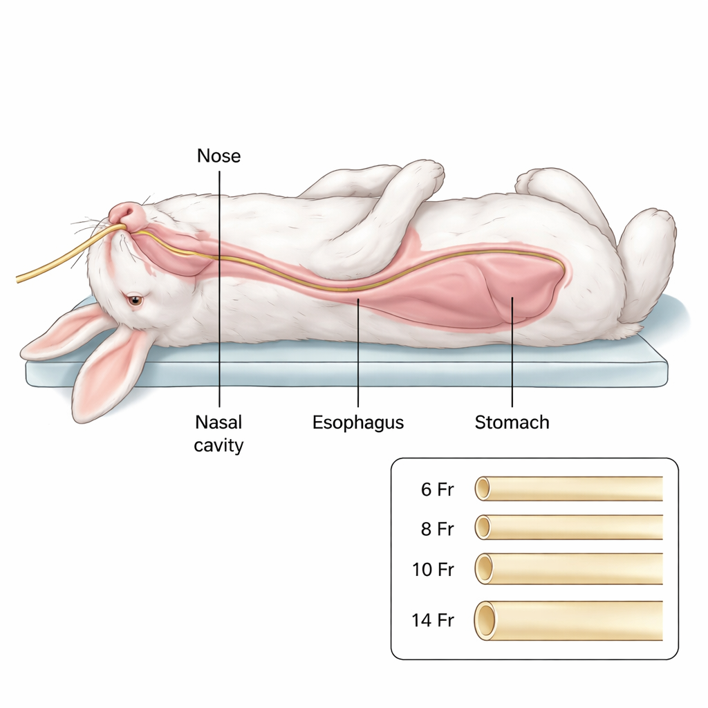
The researchers worked with healthy New Zealand rabbits and inserted standard hospital feeding tubes through the nose into the stomach, closely mimicking human practice. They first tested different tube sizes to see which could be placed reliably. Only the thinnest tube, a size called 6 French (about 2 millimeters wide), could be passed smoothly in all rabbits; larger tubes often failed or took much longer to insert. With this size chosen, the team then left the tube in place for varying times—from immediately removed up to three days—to mirror short-term use in human patients.
What happens inside the nose
Using a tiny camera called a laryngoscope, the scientists watched how the inside of the nose and throat changed over time. In animals without a tube, the lining looked smooth and healthy. Once the tube was inserted, however, distinct patterns of damage emerged. The nasal septum—the central wall that divides the nostrils—was hit hardest, showing pronounced redness, small bleeding spots, and peeling of surface cells. The curved side regions (the nasal conchae) became swollen and congested, sometimes with scattered ulcers. Farther back, in the nasopharynx, the damage was milder and appeared mainly after a day or more, and the region near the voice box (the epiglottis) was largely spared.
From surface damage to deep irritation
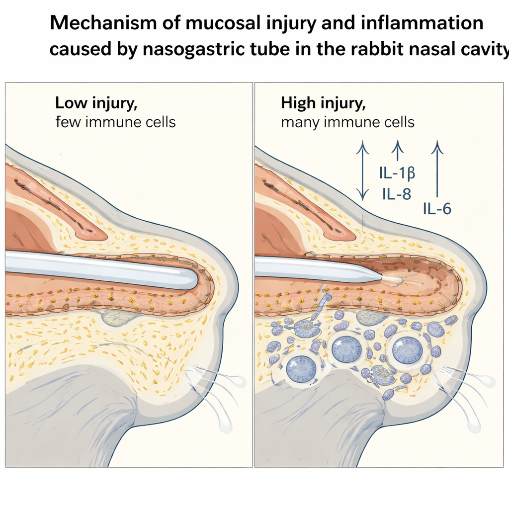
To look beyond the surface, the team took thin tissue slices from the nose and stained them for viewing under the microscope. Compared with normal animals, tube-exposed rabbits showed clear structural disruption: the top layer of cells was rubbed off, blood vessels were engorged, and many immune cells had moved into the tissue. These included neutrophils and lymphocytes, classic signs that the body was responding to injury. The longer the tube stayed in place—especially beyond 24 hours—the more intense this cellular invasion became, and the nasal septum again showed the worst changes.
Chemical signals of inflammation
The study also measured inflammatory messenger molecules that the body releases when tissue is hurt. In the damaged nasal lining, three key signaling proteins—IL-1β, IL-8, and IL-6—rose sharply after a day or more of tube placement. IL-1β is known for kicking off inflammatory cascades, IL-8 helps recruit more neutrophils to the site, and IL-6 amplifies and sustains the response. Both microscopic imaging and genetic tests showed that levels of these molecules climbed steadily with time, matching the increasing tissue damage seen under the scope.
What this means for patient comfort and safety
To a non-specialist, the message is straightforward: even a thin feeding tube can scrape and irritate the nose, and the longer it stays in, the more the tissue becomes inflamed. By establishing a carefully measured rabbit model that reproduces these changes, the researchers provide a powerful tool for testing softer materials, gentler tube designs, and improved insertion methods—without first experimenting on people. In the long run, this work may help transform one of the hospital’s most dreaded procedures into a safer, less painful experience for patients who depend on nasal feeding tubes.
Citation: Liao, X., Wang, ZG., Liu, YW. et al. A rabbit model of clinically relevant mucosal injury induced by nasogastric tube intubation. Sci Rep 16, 6810 (2026). https://doi.org/10.1038/s41598-026-36598-9
Keywords: nasogastric tube, mucosal injury, inflammation, animal model, feeding tube complications