Clear Sky Science · en
CT image-derived radiomics predicts molecular subtypes in bladder urothelial carcinoma: validation of a non-invasive classification strategy
Seeing Cancer Clues Hidden in Routine Scans
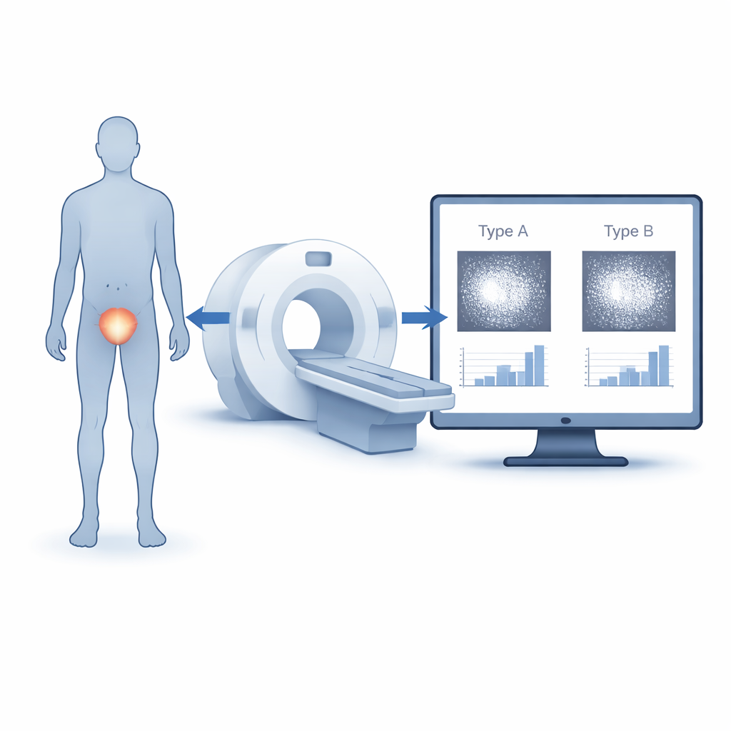
Bladder cancer is common and often requires repeated, uncomfortable procedures to understand how aggressive a patient’s tumor is. This study explores whether information already hidden inside ordinary CT scans can help doctors sort bladder tumors into biological “personalities” that respond differently to treatment—without extra needles, scopes, or costly genetic tests.
Why Tumor Personality Matters
Doctors now know that bladder cancers are not all alike. Many tumors fall into two broad molecular groups often called luminal and basal. These groups behave differently: some grow slowly and respond well to standard therapies, while others are more aggressive and may need stronger or targeted treatment. Today, assigning a tumor to one of these groups usually depends on examining tissue samples with special stains or advanced genetic tools, which are invasive, time‑consuming, and not always available. A simple, non-invasive method to infer the same information from images that patients already receive would be a major step toward more personalized care.
Turning Pictures into Numbers
The researchers focused on a technique called radiomics, which converts medical images into large sets of measurable features. Instead of a radiologist just looking at a CT scan and describing a tumor as "bright" or "irregular," radiomics measures the exact distribution and variation of pixel intensities within the mass. In this study, 96 patients with bladder urothelial carcinoma underwent contrast-enhanced CT scans before surgery. For each tumor, specialists carefully outlined the solid portion on the CT images, excluding blood, calcifications, and cystic areas, to create a precise region of interest for analysis. From these outlined areas, the team calculated basic texture measures such as average brightness, variability, and a statistic called entropy, which captures how complex or disordered the gray-scale pattern appears.
Matching Images to Molecular Types
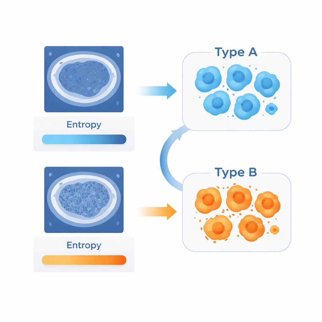
To know each tumor’s biological group, pathologists examined the removed tissue with a panel of four markers that tend to be high or low in luminal versus basal cancers. By combining the scores of these stains, tumors were classified into luminal or basal subtypes. The researchers then compared the CT-derived numbers between these groups. They found that basal tumors—which in this study were more likely to be higher grade and to invade more deeply into the bladder wall—showed both higher average brightness and, more strikingly, higher entropy on CT. In other words, the more aggressive tumors tended to look more texturally complex at the pixel level, even if that difference would not be obvious to the naked eye.
How Well Do These Image Clues Work?
To test how reliably the CT features could tell one tumor group from the other, the team used statistical tools commonly applied in diagnostic research. Among all the measured features, entropy stood out as the best single indicator of molecular subtype. Using an optimal cutoff value, entropy achieved an area under the curve of 0.79, suggesting a solid ability to distinguish basal from luminal cancers in this sample. Average brightness showed only moderate performance. Although these numbers fall short of perfection, they provide early evidence that a simple measure of image complexity could serve as a non-invasive marker of tumor biology.
What This Could Mean for Patients
This work suggests that future bladder cancer care might draw not only on what surgeons remove and pathologists stain, but also on what detailed computer analysis can reveal from routine scans. If validated in larger, multi-center studies, CT-based radiomic measures—especially entropy—could help flag more aggressive tumor types before surgery, guiding decisions about treatment intensity and follow-up. For patients, that could eventually mean more tailored therapies and fewer invasive tests, using information that is already sitting inside their imaging data.
Citation: Zhang, Q., Guo, Y., Lin, F. et al. CT image-derived radiomics predicts molecular subtypes in bladder urothelial carcinoma: validation of a non-invasive classification strategy. Sci Rep 16, 6016 (2026). https://doi.org/10.1038/s41598-026-36583-2
Keywords: bladder cancer, radiomics, CT imaging, tumor subtypes, noninvasive diagnosis