Clear Sky Science · en
The metabolites for muscle and osteoclast activity are indicators of femoral neck osteoporosis
Why hip bone health matters as we age
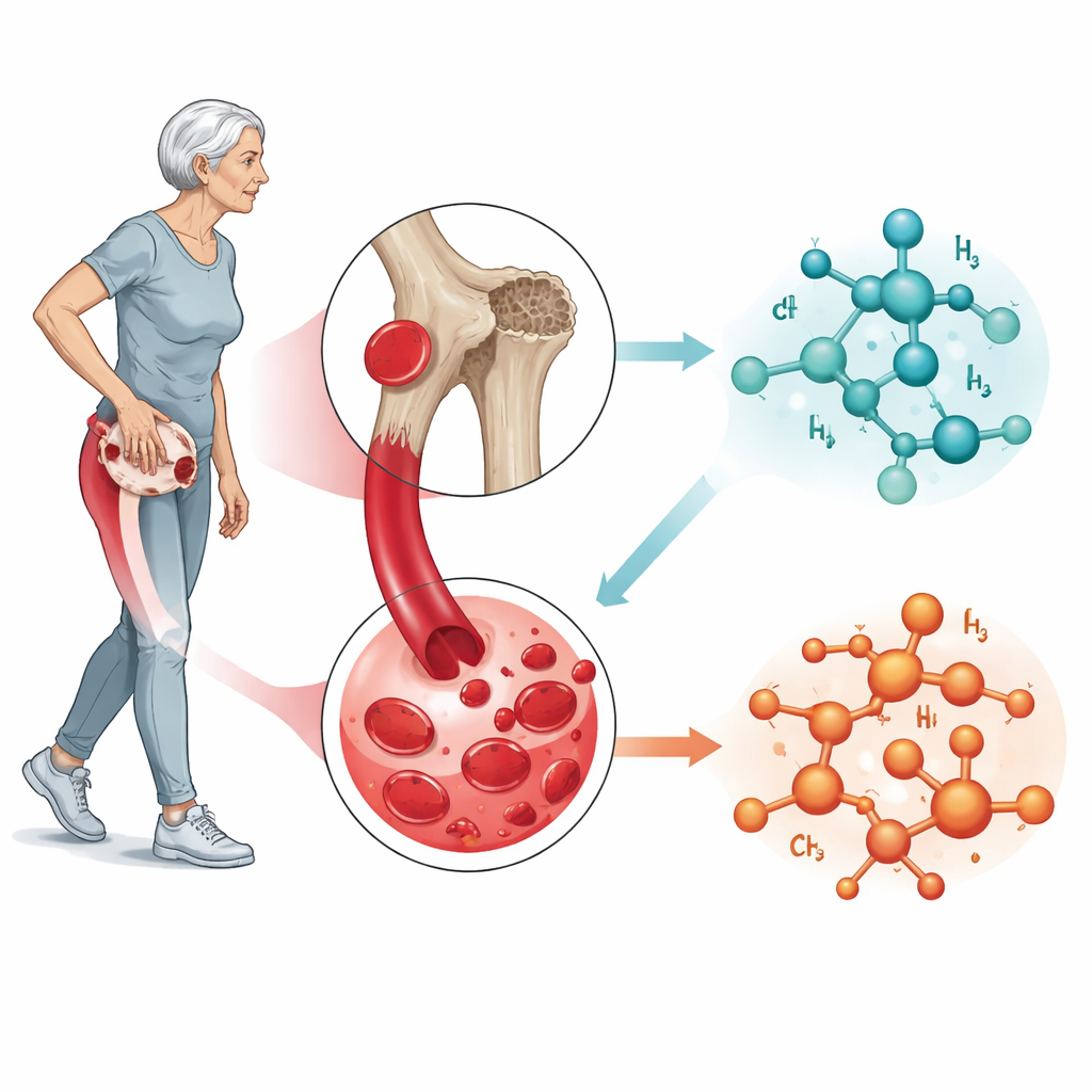
For many older adults, the difference between living independently and becoming bedridden can come down to a single fall that breaks the hip. The narrow section of the thigh bone near the hip joint—the femoral neck—is especially vulnerable. This study asks a simple but powerful question: can a routine blood test reveal early chemical signs that the femoral neck is weakening, long before a fracture occurs? By looking closely at tiny molecules circulating in the blood, the researchers uncovered a surprising chemical link between leg muscles, bone strength, and the cells that dissolve bone.
Looking inside blood for early warning signs
To explore this question, the team studied 17 community-dwelling older women in Japan, all living at home and able to visit a university hospital clinic. Using a precise X-ray technique, they measured bone density at the femoral neck and divided participants into two groups: those with osteoporosis in this region and those without. They also measured leg muscle mass, grip strength, walking ability, frailty, and memory. At the same visit, they collected whole blood—not just the liquid part—to capture a wide range of small molecules and analyzed 129 different metabolites using high-resolution mass spectrometry.
Muscle chemistry and hip bone strength go hand in hand
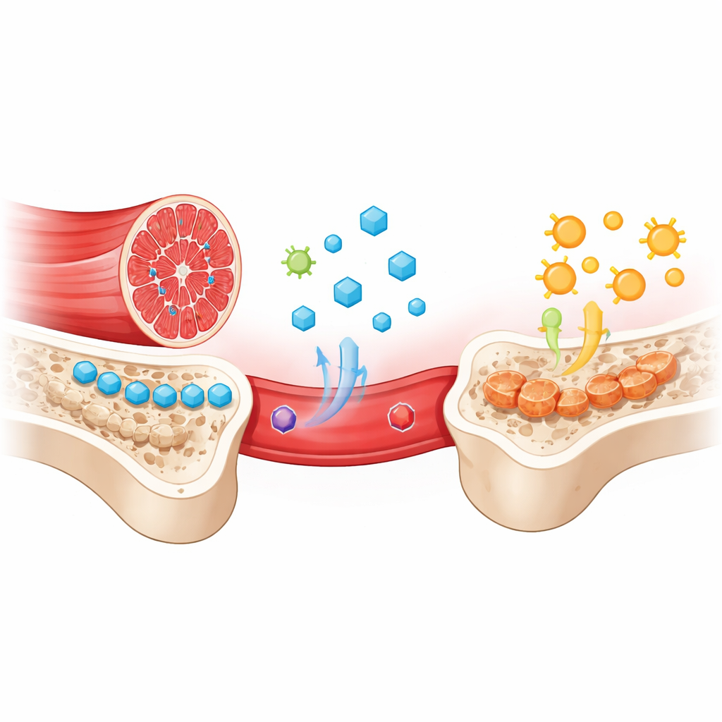
The first clear pattern was mechanical: women with femoral neck osteoporosis had noticeably less leg muscle mass and lower skeletal muscle index than those without osteoporosis. Bone density at the hip strongly tracked with leg muscle size, reinforcing the idea that bone and muscle form a single functional unit—what affects one often affects the other. When the researchers looked at blood chemistry, they found that four metabolites were significantly lower in women with femoral neck osteoporosis: phosphocreatine, malate, succinate, and histidine. The first three are closely tied to how muscles store and generate energy, particularly during activity, while histidine is connected to both muscle and antioxidant defenses. Levels of these muscle-linked metabolites rose and fell together and were higher in people with better bone density.
Chemical clues that bone-dissolving cells are active
In contrast, two other metabolites were higher in women with femoral neck osteoporosis: N1-methyladenosine and S-adenosylmethionine. Both are involved in “methylation,” a chemical tagging process that fine-tunes how genes and proteins behave inside cells. Earlier work in animals has shown that when bone-resorbing cells, called osteoclasts, become overactive, methylation pathways inside them ramp up. The elevated levels of these two methylation-related metabolites, together with reduced muscle energy markers, suggest a shift in the internal chemistry of bone and blood toward stronger osteoclast activity and bone breakdown at the femoral neck.
A chemical fingerprint that separates fragile from stronger hips
When the researchers combined all six key metabolites—four linked to muscle energy and one-antioxidant pathway, plus two linked to methylation—into a single statistical model, the pattern was striking. A standard method called principal component analysis cleanly separated women with femoral neck osteoporosis from those without, based solely on these six blood markers. The study also noted that phosphocreatine levels were lower in participants who walked more slowly in a simple chair-and-walk test that predicts fall risk, hinting that this single metabolite may reflect both muscle function and fracture risk. Although the study was small and limited to older women, its focused design and rigorous measurements make the chemical signal it uncovered particularly intriguing.
What this means for everyday health
Put simply, the study suggests that weakening hip bones leave a trace in the blood: a drop in muscle-related energy metabolites and a rise in methylation-related compounds that likely mirror overactive bone-dissolving cells. These six molecules together form a potential early-warning fingerprint of femoral neck osteoporosis, one that might someday help doctors identify high-risk patients before a fracture occurs. The findings also strengthen a practical message: keeping leg muscles strong and active is not just about movement—it may also help maintain the chemical balance that protects hip bones. While larger and more diverse studies are needed, this work opens a promising path toward blood-based tests and targeted prevention strategies to reduce the chances of life-changing hip fractures in older age.
Citation: Kameda, M., Yanagida, M. & Kondoh, H. The metabolites for muscle and osteoclast activity are indicators of femoral neck osteoporosis. Sci Rep 16, 8540 (2026). https://doi.org/10.1038/s41598-026-36570-7
Keywords: osteoporosis, hip fracture, muscle loss, bone metabolism, metabolomics