Clear Sky Science · en
High-frequency ultrasonography in quantifying paraspinal muscle remodeling after Schroth therapy for adolescent idiopathic scoliosis: a retrospective observational study
Why back muscle balance matters for growing teens
For many families, a diagnosis of adolescent idiopathic scoliosis—an unexplained sideways and twisting curve of the spine—raises urgent questions: Will the curve get worse, and can exercise really help? This study looks inside the back muscles of teenagers with scoliosis to see how a popular exercise program, known as Schroth therapy, may gently reshape those muscles over just three months, using a safe imaging method that avoids X‑rays.
Seeing scoliosis beyond the X‑ray
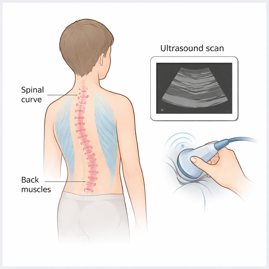
Doctors traditionally track scoliosis with standing spine X‑rays, measuring the bend with a value called the Cobb angle. While useful, repeated X‑rays expose adolescents to radiation and reveal little about the soft tissues that actually help hold the spine in place. Earlier research has shown that in scoliosis the muscles running along the spine are often out of balance—typically thinner on the outer, bulging side of the curve and bulkier on the inner, compressed side. These differences may influence how the curve behaves over time, but standard imaging tools cannot easily measure them in detail or at frequent checkups.
A closer, safer look with high‑frequency ultrasound
In this retrospective study, 50 boys and girls aged 10 to 18 with mild to moderate scoliosis completed a 12‑week program of supervised Schroth exercises, three times a week. Researchers used high‑frequency musculoskeletal ultrasound—a type of imaging that uses sound waves instead of radiation—to scan the back muscles on both sides of the spine at three key levels: the upper end, the most curved middle point, and the lower end of each person’s spinal curve. They carefully measured the width, depth, outline, and cross‑sectional area of the muscles before and after the exercise program to see whether their size and shape changed.
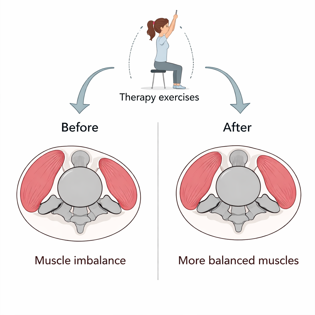
How targeted exercises reshaped the back muscles
Before training, the scans confirmed a familiar pattern: muscles on the outer, curved side of the spine were generally smaller, while those on the inner side tended to be larger, reflecting years of uneven loading. After 12 weeks of Schroth therapy, the picture shifted. At all three spinal levels, the outer‑side muscles became thicker and larger in cross‑section, showing signs of healthy growth. At the same time, the inner‑side muscles tended to shrink modestly. This two‑way adjustment suggests that the exercises did more than simply strengthen the back overall—they appeared to coax the muscles toward a more even balance around the spine.
Consistent changes across different curve severities
The researchers also divided participants into a mild group and a moderate‑to‑severe group based on their Cobb angles. In both groups, the outer‑side muscles at the middle of the curve clearly increased in size, while the inner‑side muscles decreased. Although the number of teens with more pronounced curves was small, limiting firm comparisons, the same general pattern of remodeling appeared regardless of starting severity. This hints that even when the spine is more curved, targeted three‑dimensional exercises may still help the surrounding muscles adapt in a helpful way.
What these short‑term changes may mean for care
To parents and teens, the key message is that Schroth therapy seems capable of nudging back muscles toward better balance in just three months, at least at the level of structure. High‑frequency ultrasound provided a radiation‑free window into these shifts, making it suitable for repeated monitoring during growth spurts when regular X‑rays are a concern. However, the study did not track long‑term spine shape, strength, or day‑to‑day function, and it lacked a comparison group that did not receive Schroth training. As a result, the work cannot yet prove that these muscle changes will slow curve progression or improve quality of life on their own. Even so, the findings support the idea that early, targeted exercise programs—paired with safe imaging tools—may give clinicians more options to guide care for adolescents living with scoliosis.
Citation: Tian, J., Ying, X., Ye, X. et al. High-frequency ultrasonography in quantifying paraspinal muscle remodeling after Schroth therapy for adolescent idiopathic scoliosis: a retrospective observational study. Sci Rep 16, 5707 (2026). https://doi.org/10.1038/s41598-026-36567-2
Keywords: adolescent idiopathic scoliosis, Schroth therapy, back muscle balance, ultrasound imaging, spinal curvature