Clear Sky Science · en
Comprehensive assessment of maxillary and mandibular miniplate insertion sites in normo- and hyperdivergent facial patterns using cone-beam computed tomography
Why your jawbones matter for modern braces
Invisible braces and clever springs can move teeth in dramatic ways, but behind many of these treatments sit tiny metal plates anchored directly to the jawbone. These “miniplates” act like internal handle points for pulling and pushing teeth without relying on headgear or perfect patient cooperation. This study asks a simple but crucial question: in different types of faces and in men versus women, where is the jawbone actually thick and strong enough to hold those plates safely?
Tiny anchors with a big job
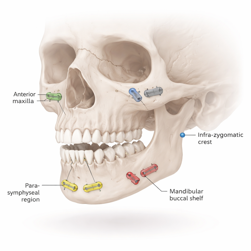
When orthodontists correct serious bite problems, they increasingly use skeletal anchorage—small titanium plates fixed to the bones of the upper and lower jaws with screws. Four regions are especially important: the front of the upper jaw, a cheekbone area above the upper molars, the front of the lower jaw, and the outer shelf of bone behind the lower molars. If the bone in these spots is too thin or too soft, plates may loosen, fail, or damage teeth. Yet until now, no single study had mapped bone thickness and bone quality for all of these regions at once, or carefully compared them between men and women and between people with different facial types.
Scanning the jaws in three dimensions
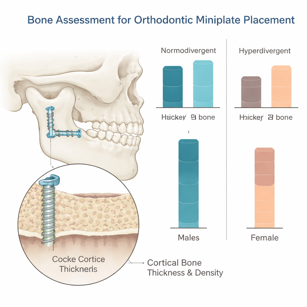
The researchers examined cone-beam CT scans—3D X‑ray images routinely used in dentistry—from 120 young adult Caucasian patients. All had full permanent teeth and no prior orthodontic or jaw surgery. The team grouped them into two facial patterns based on how steeply the lower jaw is angled: “normodivergent” (average vertical facial height) and “hyperdivergent” (longer, more open vertical faces). For each patient, they measured three key features in dozens of tiny locations in the four miniplate regions: how thick the hard outer shell of bone was, how dense that shell appeared on the scan, and, in the back of the lower jaw, how much space existed between tooth roots.
How sex and face shape change the bone landscape
Across most regions, men had thicker outer jawbone than women, especially in the front of the upper jaw and around the lower molars. An important exception was the front of the lower jaw in people with average facial proportions, where women actually had thicker bone than men. Bone density—how solid that shell looked on the scans—usually did not differ much between sexes, with a modest male advantage only in the front of the upper jaw among long‑faced patients. Facial pattern turned out to matter as much as sex: people with hyperdivergent, long faces typically showed thinner cortical bone in almost all regions than those with average vertical proportions, even when density looked similar. This suggests that their jawbones may offer less secure purchase for plates and screws, even if they appear healthy overall.
Practical guidance for placing plates
By translating these measurements into simple rules of thumb, the authors provide a roadmap for safer treatment planning. In the front of the upper jaw, thickness was limited close to the tooth roots, so they recommend placing fixation screws higher up the bone—about 16 to 20 millimeters above the gum line in women and at least 14 millimeters in men—to improve grip and avoid tooth damage. In the cheekbone region, the front of the lower jaw, and the outer shelf behind the lower molars, bone thickness and density were generally similar between sexes, though still reduced in long‑faced patients. Overall, the back lower jaw offered especially robust bone for anchoring plates in people with average facial proportions, while long‑faced women tended to have the thinnest bone in this area and may require extra caution.
What this means for future orthodontic care
For someone facing complex orthodontic treatment, these findings help explain why a clinician may insist on detailed 3D imaging and tailor the position and size of miniplates to individual anatomy rather than following a standard recipe. The study shows that sex and facial pattern subtly reshape the “landscape” of jawbone thickness, especially in the front of the upper jaw and along the lower molars. By mapping these variations, the work supports more personalized, safer placement of skeletal anchorage devices and highlights that long‑faced patients, in particular, may need more careful planning to ensure their plates stay stable throughout treatment.
Citation: Almashraqi, A.A., Sawady, M., Alamir, A.A. et al. Comprehensive assessment of maxillary and mandibular miniplate insertion sites in normo- and hyperdivergent facial patterns using cone-beam computed tomography. Sci Rep 16, 3887 (2026). https://doi.org/10.1038/s41598-026-36551-w
Keywords: orthodontic anchorage, miniplates, jaw bone thickness, cone-beam CT, facial pattern