Clear Sky Science · en
Experimental study of anterior wall sliding oblique suture technique in supermicrosurgery
Why tinier stitches could save tiny body parts
When a child’s fingertip or ear is accidentally cut off, surgeons must reconnect blood vessels thinner than a human hair to save the tissue. At this microscopic scale, even a single misplaced stitch can block blood flow and doom the reattached part. This study tested a new way of placing stitches in ultra‑small arteries in rats, asking a practical question with big implications: can a small change in how the needle enters the vessel wall make these high‑stakes operations faster, safer, and easier to learn?
A fresh twist on a standard stitch
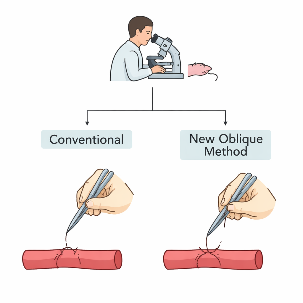
Surgeons have long relied on different ways of sewing blood vessels together, but most assume the needle goes straight down through the vessel wall. That works reasonably well for larger arteries, yet becomes risky when the vessel’s inner opening is only 0.2 millimeters wide. In such small tubes, a vertical needle can easily catch the back wall by mistake, narrowing or even closing the passageway. To solve this, the authors developed what they call the Anterior Wall Sliding Oblique Suture Technique. Instead of driving the needle straight in, the surgeon holds it at a 45–60 degree angle and gently pushes the front wall of the vessel forward before piercing it. This maneuver lets the needle pass safely in front of the back wall, reducing the chance of accidental damage.
Putting the new method to the test
To compare the new angled approach with the standard vertical one, the team operated on forty young male rats. They used the main artery in the tail, a well‑known training model for microsurgery, and created two size groups. In Group A, surgeons joined arteries about half a millimeter wide, similar to small but conventional human vessels. In Group B, they tackled arteries only 0.2 millimeters across, representing the ultra‑small targets seen in supermicrosurgery, such as tiny vessels in children’s fingertips. Within each size group, half the arteries were sewn with the traditional method and half with the new oblique technique. The researchers measured how long each repair took, how often the vessel stayed open immediately, and whether it was still open a week later.
Faster, cleaner repairs in hair‑thin vessels
The biggest advantages of the new method appeared in the smallest arteries. For vessels 0.2 millimeters wide, the angled technique cut the average sewing time by about one fifth compared with the vertical approach. More importantly, the share of arteries that were open and flowing after a single attempt jumped from 20% with the old method to 80% with the new one. A week later, only 3 out of 10 conventionally sewn arteries remained open, versus 9 out of 10 in the oblique‑stitch group. Under the microscope, the traditionally repaired microvessels often showed scarring, narrowed openings, and stitches protruding into the blood channel—features that encourage clot formation. In contrast, the oblique‑stitched vessels tended to have smoother inner linings and less injury to the outer wall.

Limits in larger vessels and what it means for surgeons
Interestingly, the new technique did not outperform the conventional one in larger, half‑millimeter arteries. Operation time was similar, and the traditional approach actually gave better long‑term flow in that size range. The authors suggest that in thicker‑walled vessels, the angled path of the needle may pull the outer layers inward, slightly twisting and narrowing the join. In everyday practice, experienced microsurgeons already achieve very high success rates with standard methods for such vessels. Where the new approach shines is at the frontier of supermicrosurgery, where tools and vessels are so small that many otherwise skilled surgeons struggle. In this study, even a relatively junior surgeon with limited experience in ultra‑small arteries could achieve high success rates once they adopted the oblique technique.
What this could mean for patients
For non‑specialists, the key takeaway is simple: by tilting the needle and letting the front wall of a tiny artery slide along it, surgeons can avoid snagging the back wall and keep the blood channel round and open. In rat tails, this small geometric change led to quicker operations and far better long‑term blood flow in hair‑thin vessels. If similar benefits hold in people, the method could make it easier to save severed fingertips and delicate parts of the face, and to perform other highly demanding reconstructive procedures. It will still need to be tested in veins, lymphatic vessels, and different connection types, but this work suggests that in supermicrosurgery, a modest shift in technique may yield outsized benefits for both surgeons and patients.
Citation: Lv, Y., Xiong, S., Ma, H. et al. Experimental study of anterior wall sliding oblique suture technique in supermicrosurgery. Sci Rep 16, 5728 (2026). https://doi.org/10.1038/s41598-026-36465-7
Keywords: supermicrosurgery, vascular anastomosis, microsurgical technique, blood vessel repair, reconstructive surgery