Clear Sky Science · en
Elevated cerebral oxygen extraction fraction in Parkinson’s disease correlates with motor impairment severity
Why brain energy matters in Parkinson’s
Parkinson’s disease is best known for its outward signs—tremor, stiffness, and slowed movement—but behind these symptoms lies a brain that is struggling to manage its energy needs. This study explores how the brains of people with early to mid-stage Parkinson’s use oxygen, a key fuel for nerve cells. By looking at how much oxygen the brain extracts from blood, the researchers hope to find a noninvasive marker that tracks how severe a person’s movement problems are, and to shed light on what is going wrong inside the affected brain regions.
Looking inside the working brain
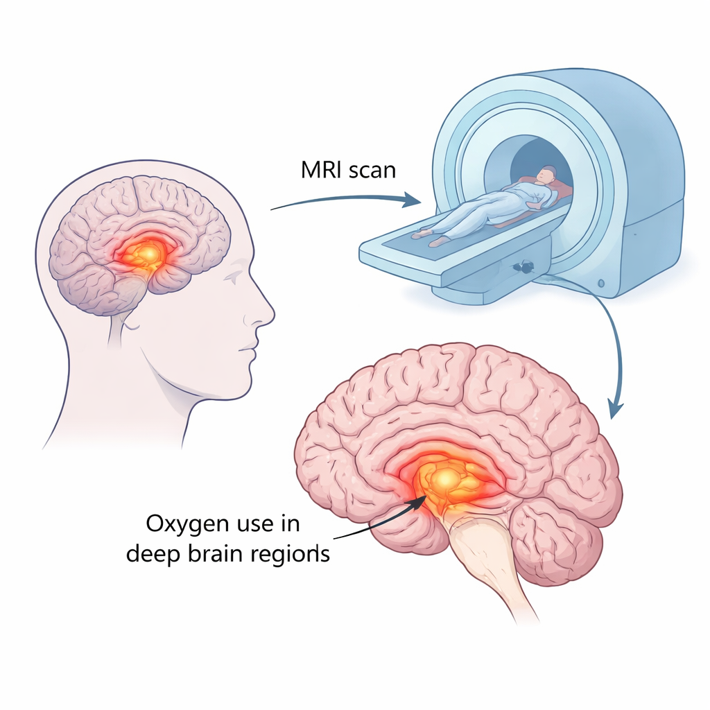
Traditionally, scientists have studied Parkinson’s by measuring how the brain uses sugar, or by using radioactive tracers to track oxygen use. These methods have revealed that certain deep brain structures involved in movement, such as the basal ganglia, show unusual patterns of activity in Parkinson’s. However, oxygen use itself has been harder to measure safely and routinely because classic techniques require short-lived radioactive substances and complex equipment. In this work, the team instead relied on advanced MRI scans—techniques already available in many hospitals—to estimate how much oxygen the brain pulls out of the blood, a quantity called the oxygen extraction fraction, or OEF. Higher OEF means brain tissue is taking up more oxygen from the same blood supply.
How the study was done
The researchers examined 50 people with Parkinson’s disease and 30 healthy volunteers. All participants underwent a specialized MRI scan that allowed the team to build maps of OEF across the whole brain. The scientists paid particular attention to the basal ganglia—regions such as the substantia nigra, red nucleus, globus pallidus, putamen, and caudate nucleus—which are central to controlling movement and are known to be disrupted in Parkinson’s. They also looked at white matter, the brain’s wiring that connects different regions. For patients, standard clinical scores of movement problems and disease stage were collected, so that brain measurements could be compared directly with symptom severity.
Where oxygen use is higher in Parkinson’s
The MRI maps revealed that people with Parkinson’s had, on average, about 8 percent higher OEF in key movement-related regions than healthy volunteers. This increase was most clearly seen in the substantia nigra, red nucleus, globus pallidus, and putamen, with somewhat weaker but still meaningful changes in the caudate nucleus and white matter. In other words, the deep hubs that help coordinate smooth motion appeared to be working harder—or at least drawing more oxygen from the blood—than in people without the disease. When the team examined the brain one tiny volume at a time across the whole head, they saw widespread pockets where OEF was elevated, especially within deep gray matter and surrounding tissue.
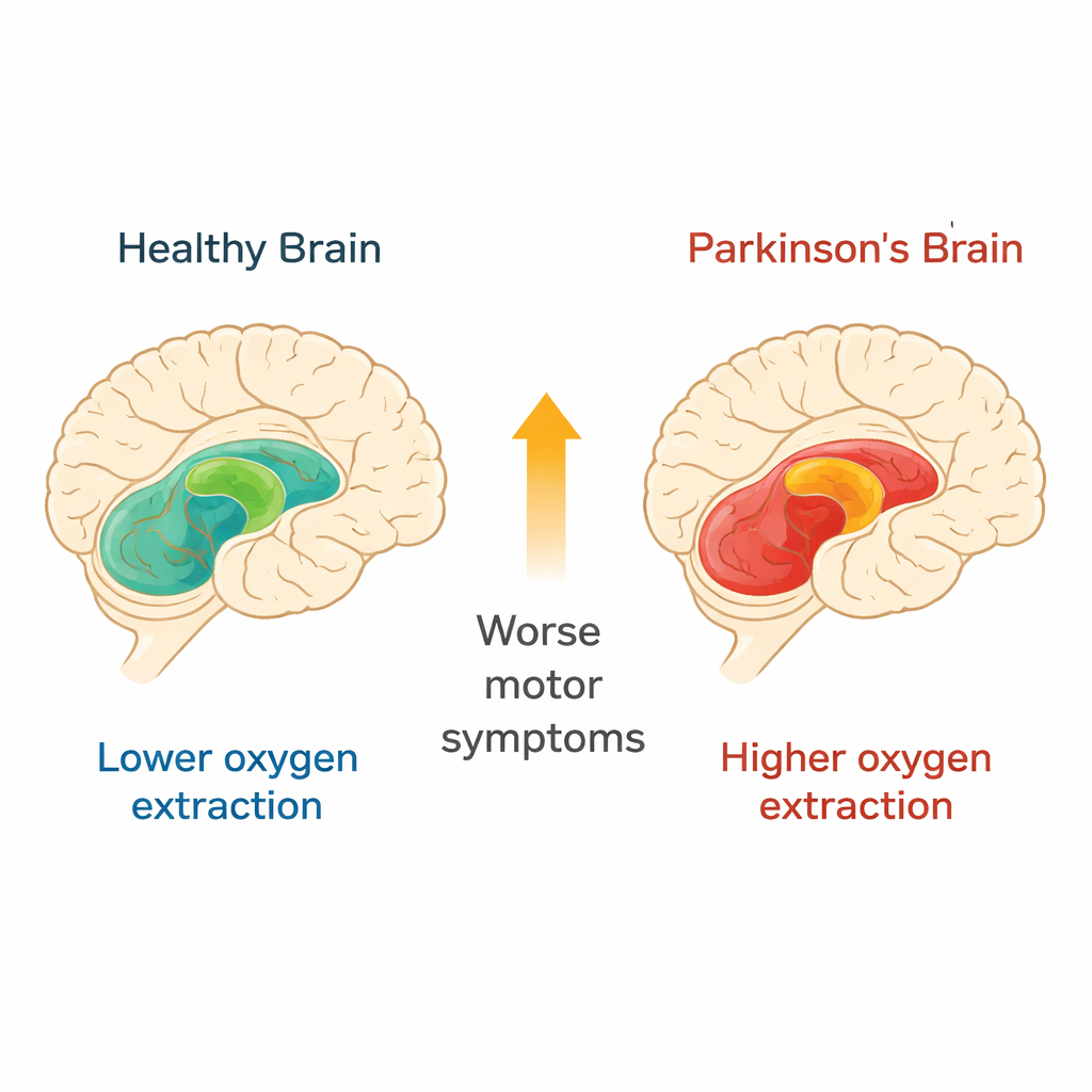
Linking brain oxygen use to movement problems
Beyond simple group differences, OEF levels tracked how impaired patients were. Higher OEF in the substantia nigra, red nucleus, globus pallidus, and white matter was associated with worse scores on a standard movement rating scale. For every 10-point increase in motor symptom score, OEF rose by roughly 1.6 percent in these regions. This pattern held even after accounting for age, and it echoed earlier findings that blood flow to the same structures also rises as symptoms worsen. Together, these results suggest that the affected circuits may be in a state of metabolic strain—either compensating for lost nerve cells by working harder, or reflecting changes in how blood vessels supply these areas.
What this means for patients and the future
The study shows that a relatively simple MRI-based measure—how much oxygen the brain extracts from the blood—rises in the deep movement centers of people with early to mid-stage Parkinson’s and increases with the severity of their motor symptoms. While OEF alone is not accurate enough to diagnose Parkinson’s on an individual basis, it offers a promising window into how the disease disrupts brain energy use. With further refinement, and by combining it with other measures such as blood flow and structural changes, OEF mapping could become a tool to monitor disease progression, test new treatments, and better understand why brain cells in Parkinson’s become so vulnerable in the first place.
Citation: Candan, H.E., Lee, D., Lee, H. et al. Elevated cerebral oxygen extraction fraction in Parkinson’s disease correlates with motor impairment severity. Sci Rep 16, 5673 (2026). https://doi.org/10.1038/s41598-026-36435-z
Keywords: Parkinson’s disease, brain oxygen use, MRI biomarkers, motor symptoms, basal ganglia