Clear Sky Science · en
Reciprocal cooperative gating fusion of SqueezeNet and ShuffleNetV2 for breast cancer detection in histopathology images
Smarter Help for Breast Cancer Diagnosis
When a lump is found in the breast, doctors often rely on tiny slices of tissue, stained and viewed under a microscope, to decide whether the cells are harmless or cancerous. This careful inspection is the gold standard for diagnosis, but it is slow, labor‑intensive, and can vary from one specialist to another. The study described here presents a computer tool that analyzes these microscope images using artificial intelligence, aiming to support pathologists with fast, consistent, and highly accurate second opinions while keeping the computing cost low enough for everyday hospital use.
The Challenge of Reading Tissue Pictures
Breast cancer is one of the most common cancers worldwide, responsible for hundreds of thousands of deaths each year. The key to better outcomes is catching and characterizing tumors early, which still depends mainly on histopathology: examining colored tissue slices under a microscope. But many tissue patterns look confusingly alike. Normal and benign (non‑dangerous) samples can share similar structures, while early‑stage and invasive cancers may blur into one another. On top of this, differences in dyes, lighting, and image contrast from lab to lab can make the same disease look quite different. Traditional computer programs struggle with these subtleties, and many modern deep‑learning systems that do better are so large and power‑hungry that they are hard to deploy outside top research centers.
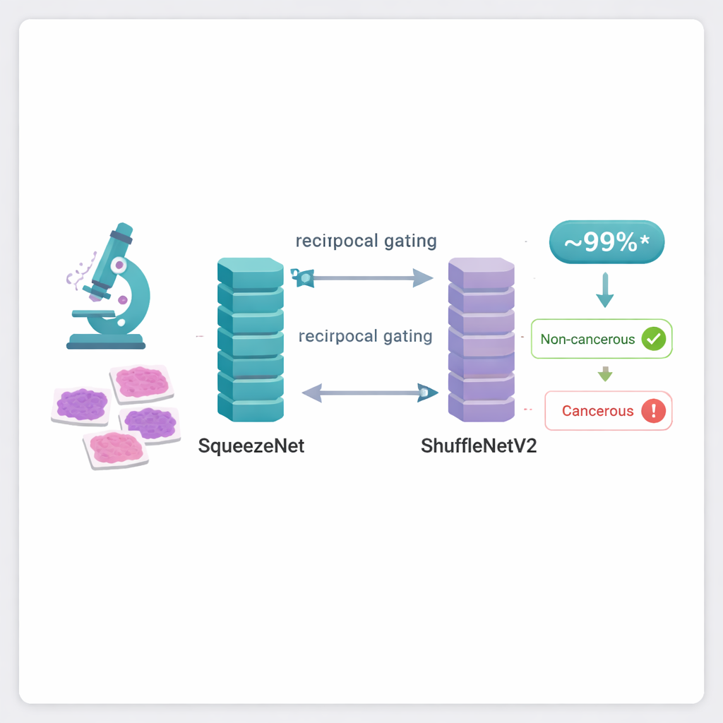
Two Compact Networks Working as a Team
To tackle this problem, the authors combine two existing compact neural networks, known as SqueezeNet and ShuffleNetV2. These models were originally designed to recognize everyday objects in photos while using far fewer calculations than heavyweight systems such as Transformers or very deep networks. In this work, they are retrained to recognize patterns in breast tissue images. Each network looks at the same microscopic picture and learns to pick out different visual clues—subtle variations in cell shapes, tissue textures, and color arrangements. On their own, each model performs well, but the real innovation is how their strengths are merged.
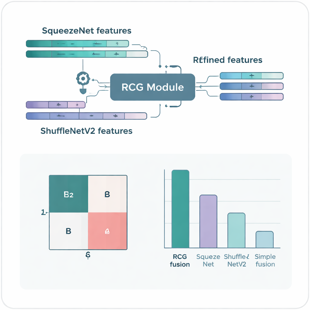
A Gating Mechanism That Filters Noise
The core idea of the study is a new "reciprocal cooperative gating" module that lets the two networks talk to each other and decide which parts of their internal signals are truly useful. Instead of simply stacking or averaging their outputs, the gating system measures how much information each channel carries and how redundant it is with what the partner network has already seen. Channels that add new, helpful detail are amplified, while those that repeat or add noise are toned down. This back‑and‑forth adjustment happens in both directions, so SqueezeNet sharpens ShuffleNetV2’s features and vice versa. The result is a single, fused representation of each image that is richer, cleaner, and more focused on the tiny differences that separate one diagnosis from another.
High Accuracy with Lightweight Computing
When tested on two widely used breast cancer image collections, ICIAR‑2018 and BreakHis, the fused system shows impressive performance. On the four‑class task—distinguishing normal tissue, benign changes, early in‑place cancer, and invasive cancer—it correctly classifies 97 percent of images. When the task is simplified to separating cancerous from non‑cancerous tissue, accuracy rises to 99 percent on ICIAR‑2018 and about 99.7 percent on BreakHis. These scores are higher than many more complex methods reported in the literature, despite the new model using relatively few parameters and modest computing power, which translates into faster predictions and lower hardware requirements.
What This Means for Patients and Clinics
In plain terms, the authors have built a lean but powerful assistant that can sift through breast tissue microscope images and label them with near‑expert reliability. Because the design is efficient, it is better suited to real‑world settings such as regional hospitals or screening centers that may not have high‑end servers. While the system does not replace pathologists, it can highlight suspicious regions, provide consistent second opinions, and potentially speed up diagnosis. The work also points to a broader trend: by cleverly combining smaller AI models and teaching them to cooperate rather than compete, it is possible to get top‑tier accuracy without the heavy cost and complexity that often stand in the way of practical medical AI.
Citation: Khati, B., Mukherjee, S., Sinitca, A. et al. Reciprocal cooperative gating fusion of SqueezeNet and ShuffleNetV2 for breast cancer detection in histopathology images. Sci Rep 16, 5904 (2026). https://doi.org/10.1038/s41598-026-36375-8
Keywords: breast cancer, histopathology, medical imaging AI, lightweight CNN, computer-aided diagnosis