Clear Sky Science · en
A comparative evaluation of parafoveal and perifoveal macular vessel density loss in glaucoma using 3 × 3 mm OCTA scans
Why tiny blood vessels in the eye matter
Glaucoma is one of the leading causes of permanent blindness, yet it often creeps in silently until much of a person’s eyesight is already lost. This study asks a deceptively simple question with big consequences for early detection: when we look at the fine network of blood vessels in the back of the eye, are we searching in the right place? By comparing two neighboring regions of the central retina, the researchers show that the outer ring of the macula – an area usually ignored in standard scans – may hold the clearest early warning signs of glaucoma.
A closer look at glaucoma and the macula
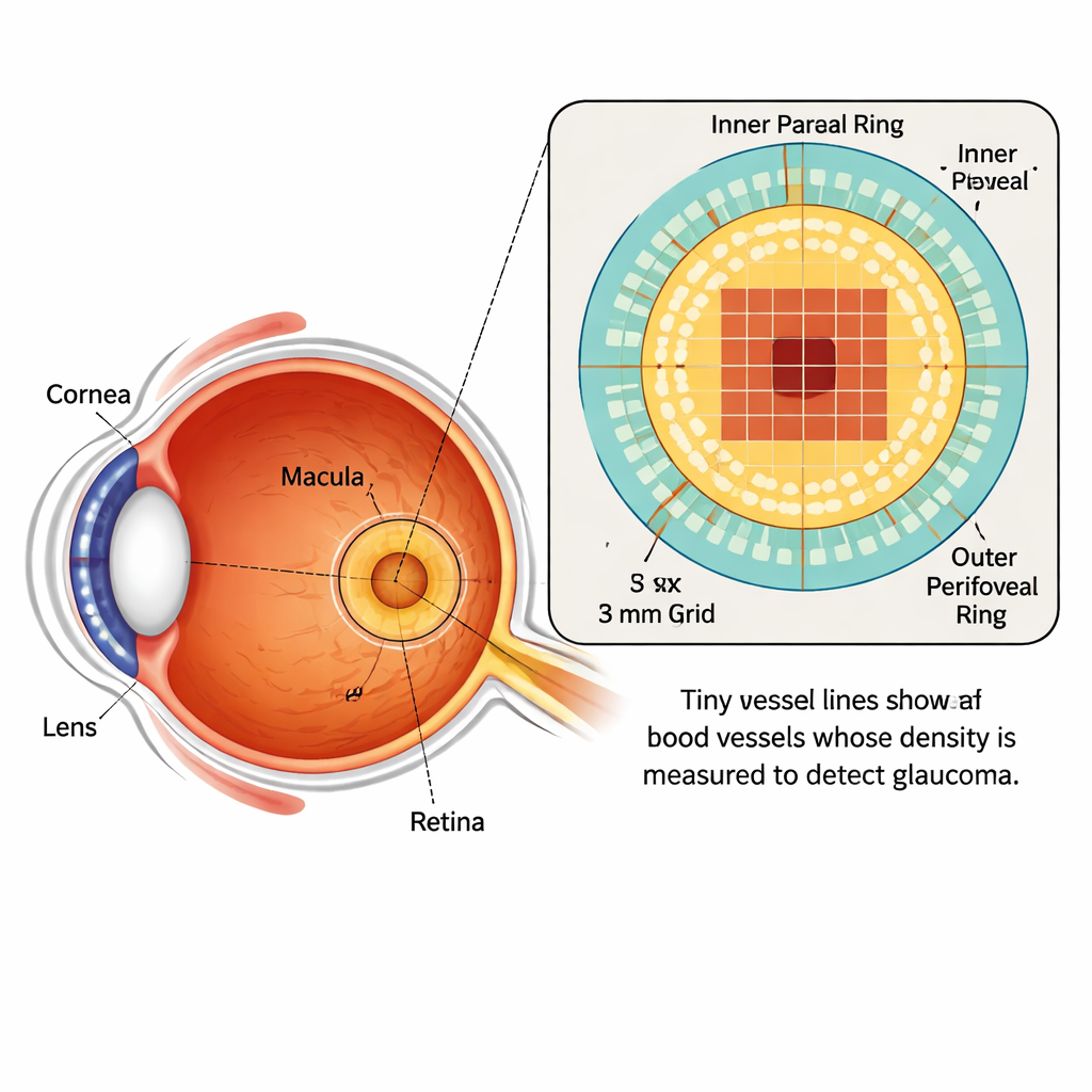
Glaucoma damages nerve cells that carry visual information from the eye to the brain. These cells, called retinal ganglion cells, are packed densely in the macula, the central part of the retina that gives us sharp, detailed vision. The macula is organized like a bull’s-eye: at the center lies the fovea, a tiny pit with no blood vessels; around it is the parafovea, and farther out is the perifovea. Modern imaging techniques can now map both the structure and blood flow in these layers without touching the eye, offering a way to spot disease before people notice vision loss.
Measuring the eye’s micro–plumbing
The team used optical coherence tomography angiography (OCTA), a noninvasive scan that captures moving blood cells in the retina’s smallest vessels. In 352 eyes (198 with glaucoma and 154 healthy), they measured “vessel density” – the fraction of each region occupied by blood vessels – in three layers of the retinal circulation. These layers are known as the superficial vascular plexus, the intermediate capillary plexus, and the deep capillary plexus. Instead of relying on rough averages, the researchers divided the inner parafoveal ring into 12 slices and the outer perifoveal region into four quadrants, then used computer algorithms called support vector machines to learn how well these patterns could distinguish diseased from healthy eyes.
Outer ring beats inner ring
When the researchers compared how accurately their models separated glaucoma from normal eyes, the perifoveal region consistently outperformed the parafoveal region in all three vessel layers. The clearest advantage appeared in the superficial layer that feeds the nerve fiber layer and ganglion cells, the very structures most harmed by glaucoma. Here, the outer region produced a very high diagnostic score, meaning its vessel patterns matched glaucoma status more closely than those from the inner ring. Even in the intermediate and deep layers, where the difference was smaller, the outer area still carried more useful information. Statistical tests confirmed that many of these differences were unlikely to be due to chance.
Rethinking how we scan for glaucoma
These results challenge common practice in eye imaging. Standard 3×3 millimeter OCTA scans of the macula usually focus their vessel-density analysis on the parafovea and leave out large parts of the perifovea. Earlier work with deep learning had hinted that computer models were paying special attention to the corners and edges of these scans; this study shows that, even with simple numerical vessel measurements, those same outer areas are indeed more informative. The authors argue that the issue is less about using a bigger scan window and more about paying attention to the right zones within the images we already take.
What this means for patients
For patients and clinicians, the message is encouraging. The smaller 3×3 millimeter scans are fast and provide high detail, making them practical for everyday clinic use. By including the perifoveal region in vessel-density analysis, doctors could gain a stronger, earlier signal of glaucoma damage without changing the hardware – only the way the data are interpreted. Detecting disease sooner would allow treatment to begin earlier, potentially preserving vision for many more years. Future studies will need to test these findings across different glaucoma types and stages, and compare them directly with larger scan sizes, but this work suggests that vital clues to glaucoma may already be hiding in the outer ring of the macula.
Citation: Garcia Kahmeyer, D., Mardin, C., Lämmer, R. et al. A comparative evaluation of parafoveal and perifoveal macular vessel density loss in glaucoma using 3 × 3 mm OCTA scans. Sci Rep 16, 3051 (2026). https://doi.org/10.1038/s41598-026-36230-w
Keywords: glaucoma, macula, retinal blood vessels, OCTA imaging, early diagnosis