Clear Sky Science · en
Quantitative assessment of age-related retinal and choriocapillaris blood flow changes in a healthy Korean population using optical coherence tomography angiography
Why the Eye’s Blood Supply Matters as We Age
As we grow older, many of us worry about losing our sight to conditions like macular degeneration or diabetic eye disease. Behind these problems lies a simple question: how well is blood reaching the light‑sensing tissue at the back of the eye? This study used a cutting‑edge, noninvasive imaging method to map how blood flow in different layers of the eye changes with age in healthy Korean adults. Understanding these normal patterns is crucial for spotting early warning signs of disease.
A New Way to Look at Tiny Vessels
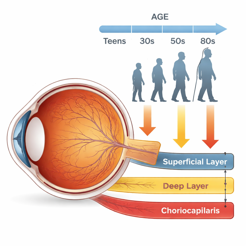
The research team relied on optical coherence tomography angiography, or OCTA, which acts like an ultrasound made of light. Instead of injecting dye into the bloodstream, the machine detects the motion of red blood cells to sketch out a detailed map of tiny vessels in the retina and just beneath it. The scientists focused on three key layers at the center of vision, the macula: a “surface” network of blood vessels, a deeper network, and a thin bed of capillaries called the choriocapillaris that nourishes the light‑sensing cells.
Carefully Measuring Healthy Eyes
To build a reliable picture of normal aging, the study included 351 healthy Korean participants, from teenagers and young adults through people in their 80s. Anyone with eye disease, major medical conditions like diabetes or high blood pressure, or poor‑quality scans was excluded. Each eye was scanned twice with a wide 6×6 millimeter pattern centered on the fovea, the point of sharpest vision. Advanced image‑processing steps were used to remove artifacts and large overlying vessels so that only the fine capillary networks remained. The researchers then calculated how much of each image was occupied by blood vessels or, in the case of the choriocapillaris, how much area showed “flow deficits,” places where blood signal was missing.
Different Layers, Different Aging Patterns
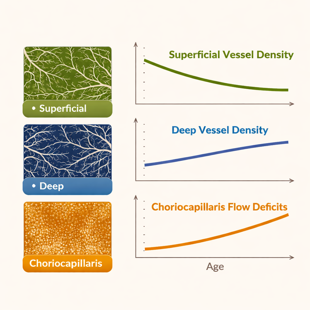
The results revealed that not all vessel layers age in the same way. In the surface layer, vessel coverage was highest in people in their 20s and stayed fairly stable through the 30s and 40s, then began a slow but clear decline starting in the 50s and continuing into the 80s. The deep layer behaved very differently: its vessel coverage was similar from the 20s through the 50s, but then gradually increased from the 60s onward. Meanwhile, the choriocapillaris showed the most striking change. Areas without detectable blood flow increased steadily with each decade, beginning as early as the 30s and rising sharply into older age, suggesting a progressive thinning or dropout of this crucial layer.
Clues to the Eye’s Coping Strategies
These patterns suggest that the aging eye may try to compensate for loss of blood flow in some regions by remodeling others. The slow decline of surface vessels, combined with the rise in deeper vessel density in later life, fits with the idea that deeper capillaries may expand or carry more flow as surface arteries stiffen and narrow with age. At the same time, the steady loss of choriocapillaris blood flow echoes earlier tissue studies that show shrinking of this layer in older adults, changes that are thought to contribute to age‑related macular degeneration. The authors also found that lens status and image quality influenced the measurements, underlining the importance of strict imaging standards when using OCTA in clinics or research.
What This Means for Protecting Vision
For a lay reader, the main takeaway is that the back of the eye does not age uniformly. Even in healthy people, the surface vessels, deeper vessels, and underlying capillary bed each follow their own trajectory over the decades. By precisely charting these normal trends in a large group of Korean adults, this study provides a reference map for eye doctors. Future patients whose scans fall outside these age‑based patterns may be flagged earlier for conditions like macular degeneration, vascular blockages, or diabetic damage. In short, knowing how a healthy eye ages gives clinicians a better chance to detect trouble early—before it steals vision.
Citation: Jeong, Y.H., Yang, S.C., Kim, T.Y. et al. Quantitative assessment of age-related retinal and choriocapillaris blood flow changes in a healthy Korean population using optical coherence tomography angiography. Sci Rep 16, 5752 (2026). https://doi.org/10.1038/s41598-026-36184-z
Keywords: retinal blood flow, aging eye, optical coherence tomography angiography, macular health, choriocapillaris