Clear Sky Science · en
In vitro experiments and bioinformatic analyses implicate KAT2A in the occurrence and development of hepatocellular carcinoma
Why this liver cancer story matters
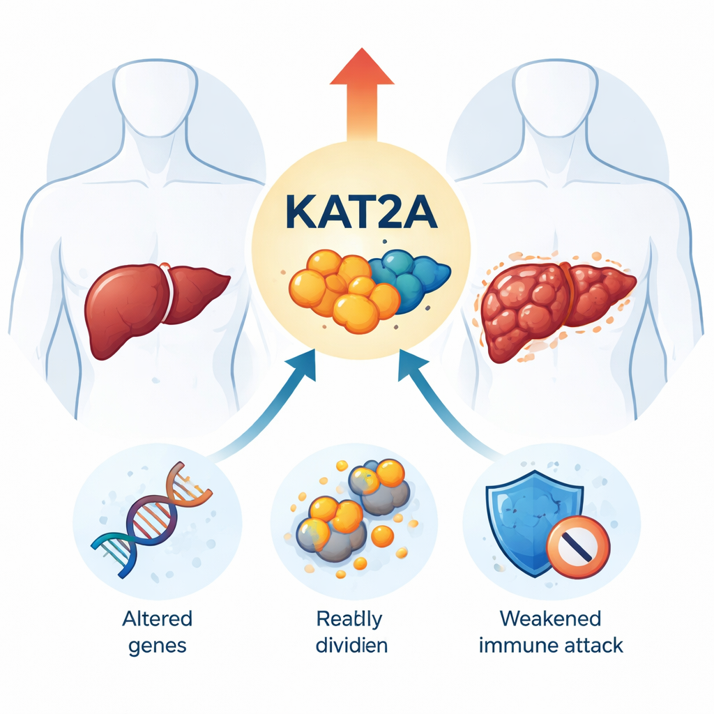
Liver cancer is one of the deadliest cancers worldwide, in large part because it is often discovered too late and doctors still lack highly reliable tests and targets for treatment. This study focuses on a little-known protein called KAT2A and asks a simple but important question: does this molecule help liver tumors grow and hide from the immune system, and could it become a new clue for diagnosis and therapy?
A closer look at a key troublemaker
KAT2A is a protein that helps control how DNA is packaged and how genes are turned on or off. In many cancers it acts like a volume knob that turns up growth signals. The researchers began by comparing KAT2A levels in thousands of tumor and healthy tissue samples stored in major international databases. They found that KAT2A is higher in many cancers, but especially in liver cancer. In liver tumors, both the gene message (mRNA) and the actual protein were clearly elevated compared with nearby normal liver tissue, and patients whose tumors had more KAT2A tended to have more advanced disease.
Linking KAT2A to patient outcomes
To understand what this means for real people, the team divided liver cancer patients into groups with high or low KAT2A levels and tracked how long they stayed free of disease progression. Those with high KAT2A had a shorter progression-free interval, meaning their cancer tended to come back or worsen sooner. Using a statistical tool called a ROC curve, the researchers showed that KAT2A levels could distinguish liver cancer tissue from normal liver with very high accuracy. Even after accounting for tumor stage and spread, KAT2A remained one of the strongest predictors of outcome, suggesting that it could serve as a powerful prognostic marker.
What KAT2A is doing inside tumor cells
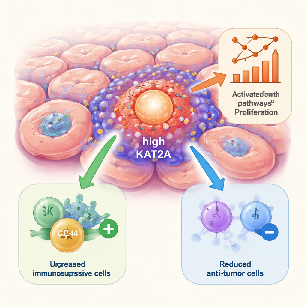
The scientists then asked what other genes change when KAT2A is high. By comparing tumors with high versus low KAT2A, they identified 125 genes whose activity shifted in a consistent way. These genes clustered in well-known cancer pathways that drive cell division, survival, and resistance to treatment, including major growth circuits often targeted by modern drugs. In laboratory experiments using human liver cancer cell lines, the team turned down KAT2A using small interfering RNA. Cells with reduced KAT2A grew more slowly, formed fewer colonies, and migrated less efficiently across a dish, all signs that KAT2A helps maintain the aggressive behavior of liver cancer cells.
How KAT2A shapes the tumor neighborhood
Cancer does not grow in isolation; it interacts constantly with immune and support cells around it. Using computational tools that infer immune cell types from tumor gene data, the researchers found that tumors with high KAT2A contained more regulatory T cells and certain dendritic cells, both associated with dampening immune attacks. At the same time, there were fewer memory B cells, neutrophils, and some macrophages that can contribute to anti-tumor defenses. Single-cell RNA sequencing, which profiles individual cells from patient tumors one by one, showed that KAT2A is especially abundant in cholangiocytes (bile duct–like cells), actively dividing cells, and dendritic cells. Communication maps between cell types suggested that dendritic cells with high KAT2A send strong signals to many other cells, potentially helping to sculpt an immunosuppressive environment.
A new axis in liver cancer biology
To explore how KAT2A is controlled, the team mined four large databases of transcription factors—the proteins that act as master switches for genes. The only factor consistently connected to KAT2A across all sources was MYC, a famous cancer-promoting protein. In liver cancer samples, KAT2A and MYC levels rose and fell together. Earlier work has shown that KAT2A can modify MYC and that MYC can stimulate many growth-related genes, suggesting a reinforcing partnership: MYC may help turn on KAT2A, while KAT2A may boost MYC’s activity, jointly pushing liver cells toward uncontrolled growth.
What this means for patients
Taken together, the findings paint KAT2A as more than a bystander: it appears to help liver tumors grow, spread, and disarm the body’s immune response. Because its levels are clearly higher in tumor tissue, strongly linked to patient prognosis, and influential in cell behavior in the lab, KAT2A stands out as a promising marker for earlier diagnosis and risk prediction. In the long run, drugs that inhibit KAT2A, possibly in combination with existing immune checkpoint therapies, could open a new front in the fight against liver cancer, though careful animal studies and clinical trials will be needed before such strategies reach the clinic.
Citation: Xu, ZY., Tan, JH., Li, JX. et al. In vitro experiments and bioinformatic analyses implicate KAT2A in the occurrence and development of hepatocellular carcinoma. Sci Rep 16, 5737 (2026). https://doi.org/10.1038/s41598-026-36174-1
Keywords: liver cancer, KAT2A, tumor microenvironment, immunotherapy, biomarkers