Clear Sky Science · en
Brain tumor classification from MRI images using a multi-scale channel attention CNN integrated with SVM
Smarter Scans for Brain Tumor Care
When doctors look at brain scans to decide whether a patient has a tumor—and what kind it is—they face a demanding, time‑critical task. This study explores a new kind of computer assistant that learns to read MRI images more accurately and consistently than many existing methods. By combining two powerful approaches from artificial intelligence, the system aims to support radiologists with faster, more reliable second opinions, potentially leading to earlier diagnosis and better treatment planning.
Why Classifying Brain Tumors Is So Hard
Brain MRIs are rich, complex images. Tumors can vary greatly in shape, size, and texture, and normal brain structures already look intricate to begin with. Human experts can disagree, especially when cases are subtle. Traditional computer programs either rely on hand‑crafted measurements or on standard deep‑learning models that do not always capture all the crucial details. These older systems may struggle with balancing sensitivity (catching real tumors) and specificity (avoiding false alarms), and can become unreliable when faced with new patients whose images differ slightly from the training data.
A Two‑Part AI That Looks at Images Many Ways
The researchers designed a hybrid system called MCACNN‑SVM that splits the job into two stages: seeing and deciding. First, a specialized deep‑learning network examines each MRI slice through several “lenses” at once—small, medium, and large viewing windows. This multi‑scale design lets the model pick up both fine edges and broader structures, such as subtle tumor borders and overall shape. A built‑in “attention” module then learns which image channels carry the most useful information and boosts those signals, while downplaying less relevant background patterns. 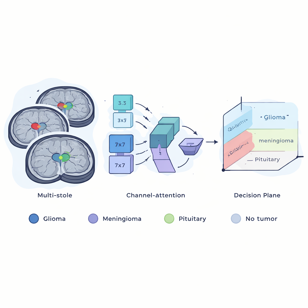
Training on Real‑World Hospital Images
To test their approach, the authors used a public dataset of more than 7,000 MRI slices collected from Chinese hospitals. Each image was already anonymized and cleaned, and labeled as one of four groups: glioma, meningioma, pituitary tumor, or no tumor. The images were resized and lightly transformed—rotated, flipped, and zoomed—to mimic the variety seen in clinical practice, helping the model avoid overfitting to a narrow set of examples. During training, the team carefully adjusted how quickly the network learned by cycling the learning rate up and down in a smooth, wave‑like pattern. This “warm restart” schedule helps the model escape poor solutions and settle into a more reliable state, while a grid search tuned the key settings of the final support vector machine so it could make the sharpest possible distinctions between tumor types.
How Well the System Performed
On unseen test images, the hybrid model correctly classified brain scans about 98% of the time, with especially strong performance in recognizing pituitary tumors and scans with no tumor at all. A detailed breakdown showed high precision and recall across categories, and near‑perfect scores on a common summary measure called the ROC‑AUC, which tracks how well the system separates positive from negative cases. 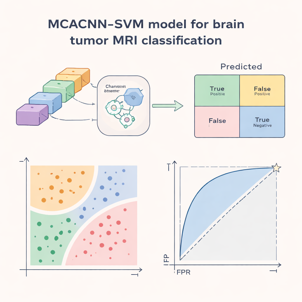
What This Could Mean for Patients
In simple terms, this work demonstrates that letting one AI specialize in “seeing” and another in “deciding” can yield a smarter assistant for reading brain MRIs. While the system is not a replacement for radiologists, it could act as a high‑quality second reader, flagging suspicious regions, helping to distinguish between tumor types, and reducing the odds of missed or incorrect diagnoses. The authors note that more testing on diverse hospitals, scanners, and image qualities is still needed, and that future versions will aim to be even lighter and more broadly applicable. Nonetheless, the study points toward AI tools that are accurate, robust, and practical enough to support real‑world brain tumor care.
Citation: Ke, L., Hu, G., Zhao, M. et al. Brain tumor classification from MRI images using a multi-scale channel attention CNN integrated with SVM. Sci Rep 16, 6297 (2026). https://doi.org/10.1038/s41598-026-36164-3
Keywords: brain tumor MRI, medical imaging AI, deep learning, support vector machine, tumor classification