Clear Sky Science · en
Scalable DICOM 3D-printed phantoms mimicking marine mammal bone and soft tissue
Why a Fake Sea Lion Matters
From ocean aquariums to rescue centers, veterinarians care for California sea lions that may be sick, injured, or poisoned by harmful algal blooms. Drawing blood from these powerful, sensitive animals is vital for diagnosis, yet hard to learn safely on living patients. This study describes how researchers turned medical scan data into a life-like, 3D-printed model—or “phantom”—of a sea lion’s hip region. The phantom feels and behaves much like real tissue, giving trainees a realistic practice tool and pointing the way to new medical models for both animals and people.
Turning Scans Into Solid Shapes
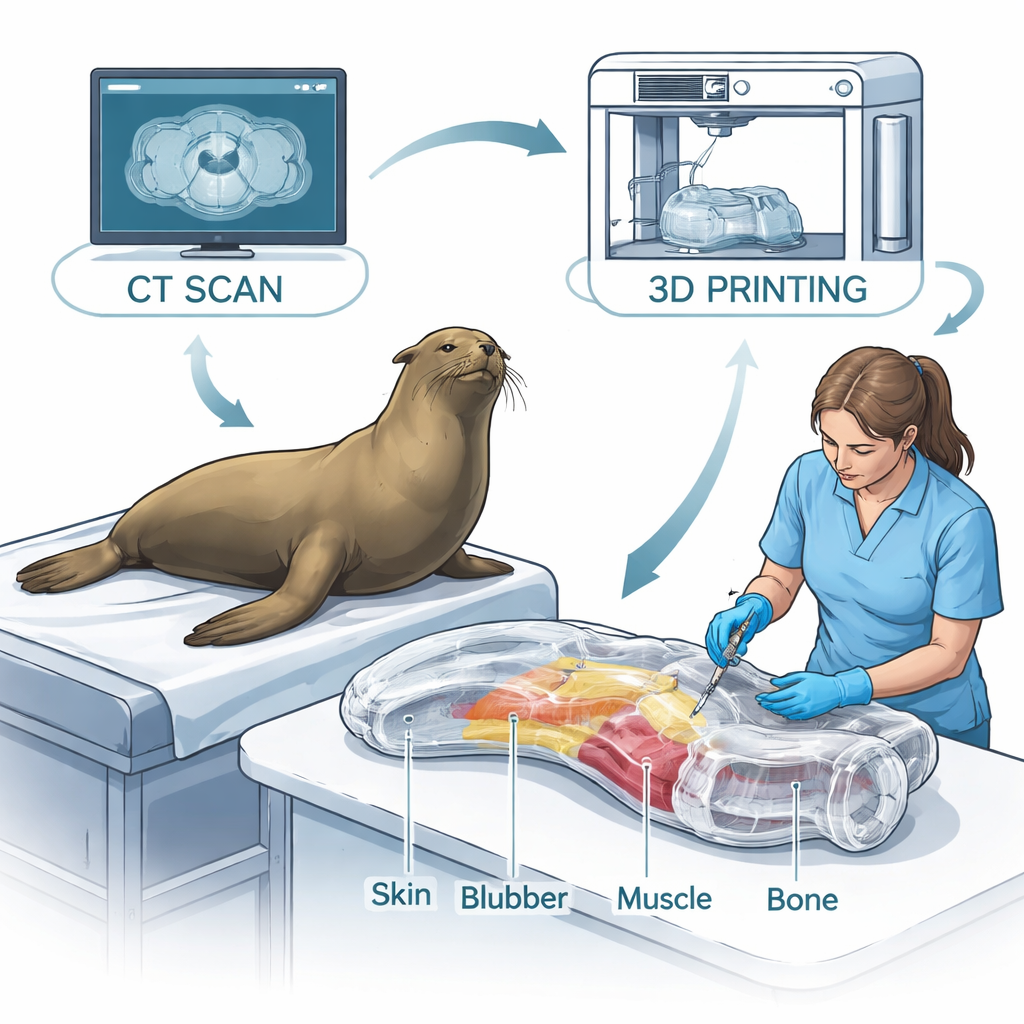
The team began with detailed CT and MRI scans of a real California sea lion, supplied by the U.S. Navy Marine Mammal Program. These scans, stored in the standard DICOM medical format, show how dense each tiny volume of tissue is, from soft blubber to hard bone. Using specialized software, the researchers "segmented" the images, separating out bones and soft tissues based on their brightness in the scans. They then cleaned and smoothed the digital models, cropped away the exam table and other clutter, and split the skeleton into practical sections such as legs, flippers, and pelvis and spine. The result was an anatomically faithful digital lower body, with special attention to the region where blood is commonly drawn, just behind the hip bones.
Building a Layered Body From the Inside Out
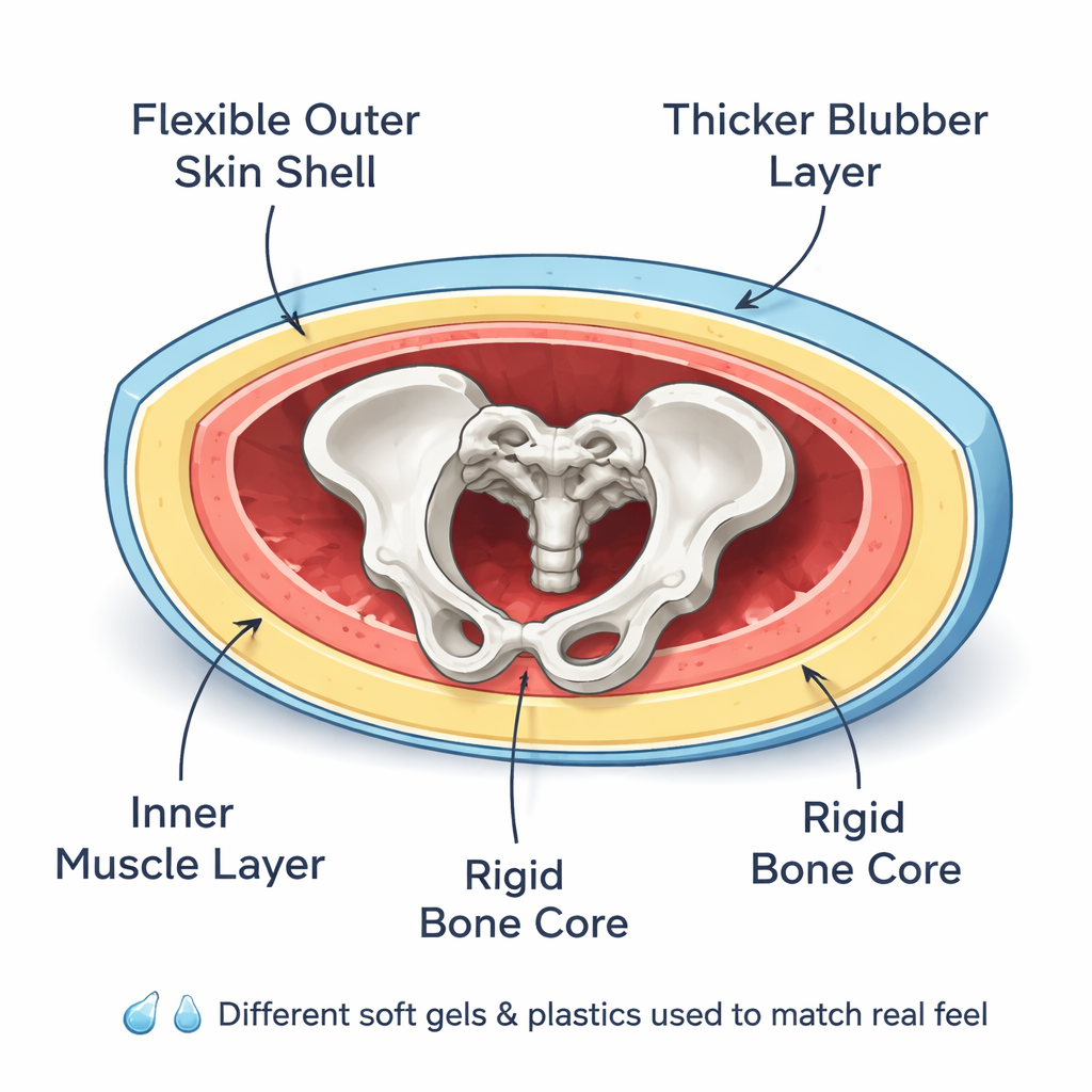
Rather than making a single solid block, the researchers designed the phantom as four distinct layers that mimic real anatomy: bone, muscle, blubber, and skin. The bone shapes were exported directly as 3D-printable files and printed at reduced scales using high-resolution stereolithography printers. A flexible outer shell was designed around the body, hollowed to create a cavity, and split open at the top so that the bones and soft tissues could be inserted. This shell does double duty: it acts as the visible “skin” of the phantom and serves as a mold for casting the inner gels. Natural bony landmarks, like tail vertebrae and flipper sockets, were preserved so that the skeleton could be aligned precisely inside the shell, reproducing the feel of real landmarks that clinicians rely on by touch.
Making Fake Tissues Feel Real
To capture how real sea lion tissues deform under a needle or a hand, the team turned to a family of clear, reusable medical gelatins. These gels come in several grades, from very firm to very soft. Using a dynamic mechanical analyzer, the researchers compressed small gel samples in a controlled way to measure their stiffness and energy loss over repeated loading, similar to pushing on and releasing living tissue. By comparing these measurements with known properties of sea lion blubber, muscle, and bone, they chose specific gels for each layer: a stiffer gel near the bone to stand in for tough connective tissue, a softer gel for muscle, and an intermediate gel for the thick blubber layer. A tough yet somewhat flexible plastic resin was selected for the skeleton, while a transparent, stretchy resin formed the outer skin, allowing the internal bones to remain visible during practice.
From Digital Model to Working Phantom
With materials and geometry set, the researchers assembled the phantom step by step. First, they printed the bones and dipped them in a firm gel to represent tendons and tightly bound muscle near joints. The clear skin shell was printed separately. Then they calculated the volume of blubber and muscle space inside the shell at different scales to know how much gel to melt and pour. Working in vacuum ovens and ice baths to control bubbles and cooling, they poured a blubber layer along the shell walls, placed the skeleton in its exact position, and finally filled the remaining space with a soft muscle gel. After a day of curing, they gently heat-polished the exposed surface to smooth it without warping the shell. The finished model closely matched the original 3D render, held together well during handling, and let users both feel and see the internal structures.
What This Means for Training and Beyond
For trainees, this sea lion phantom offers a realistic way to practice finding bony landmarks and inserting needles in the correct spot, without putting live animals at risk. Because the workflow begins with routine medical images, it can be adapted to other body regions, other species, and even human patients. The study also shows how image-based design and carefully tested soft materials can reproduce living tissues well enough for training, and potentially for soft robotic devices or custom implants. In short, the researchers have demonstrated a practical recipe for turning digital anatomy into touchable, scalable models that bring the feel of the clinic or the ocean rescue center into the lab or classroom.
Citation: Fisher, D., Minaian, N., McClain, A. et al. Scalable DICOM 3D-printed phantoms mimicking marine mammal bone and soft tissue. Sci Rep 16, 5929 (2026). https://doi.org/10.1038/s41598-026-36154-5
Keywords: 3D-printed phantom, California sea lion, veterinary training, medical imaging, tissue-mimicking gels