Clear Sky Science · en
Putative long range mossy fiber sprouting and regional cytochrome c oxidase alteration in the hippocampus of patients with mesial temporal lobe epilepsy
When Seizures Rewire the Brain’s Memory Center
Mesial temporal lobe epilepsy (MTLE) is one of the most stubborn forms of epilepsy, often resisting medication and stealing memory and independence from patients in their most productive years. This study looks deep inside the brain’s memory hub—the hippocampus—to see how seizure activity may physically rewire its circuits and change how brain cells use energy, offering clues to why seizures persist and how surgeons and future therapies might better target them.
The Brain Region at the Heart of the Problem
In MTLE, seizures usually arise from the hippocampus, a seahorse-shaped structure vital for forming new memories. Many patients develop “hippocampal sclerosis,” where key groups of neurons die and supporting cells scar. The authors examined 20 hippocampi surgically removed from people with drug-resistant MTLE and compared them with 20 donated, non-epileptic brains. They focused on the dentate gyrus (the entry gate of the hippocampus), areas called CA3, CA2, CA1, and a nearby output zone known as the subiculum. Using a panel of molecular stains, they could count surviving neurons, trace specific nerve fiber bundles called mossy fibers, and measure the activity of a major energy-producing enzyme in cell powerhouses (mitochondria).
Lost Neurons and Scrambled Cell Layers
First, the team confirmed the classic damage pattern seen in MTLE. In the surgical samples, many neurons were missing from the dentate hilus and from CA3 and CA1, with some loss extending into CA2 and the subiculum. The normally neat rows of cells in the dentate gyrus were disrupted: granule cells had spread out and even formed island-like clusters, a phenomenon known as granule cell dispersion. Two independent markers of neurons showed the same story—severe thinning of key relay stations in the hippocampal circuit, while some regions such as CA2 and parts of the subiculum were relatively spared but appeared structurally disturbed.
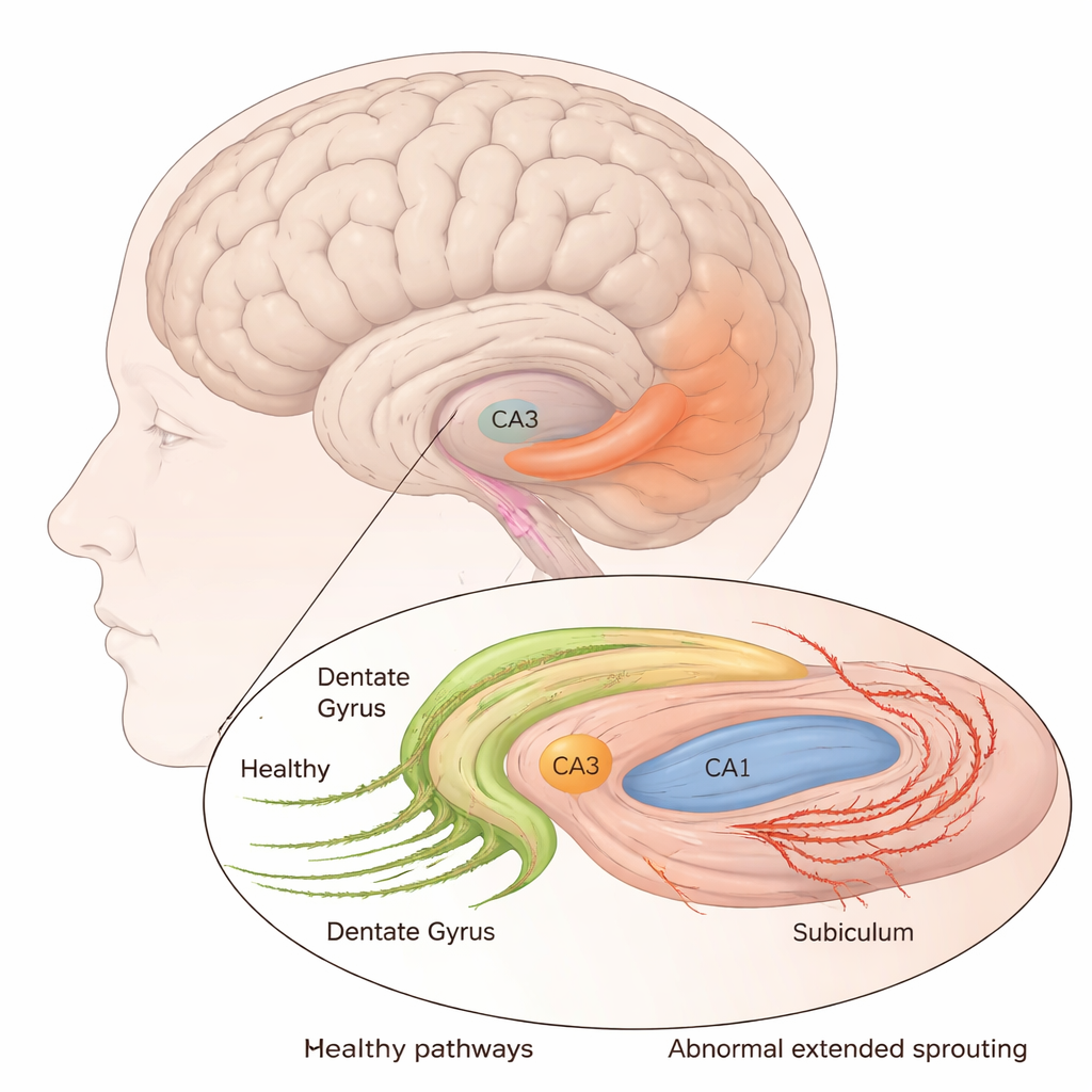
Sprouting Wires That Reach Further Than Expected
The most striking changes involved mossy fibers, the axons of dentate granule cells that normally make short-range connections within the hippocampus. Using three different markers, the researchers found that these fibers had not only regrown into a nearby layer (the inner molecular layer), a well-known feature of epilepsy, but also seemed to extend further than usual. In MTLE tissue, the inner molecular layer was widened and strongly stained, while the original mossy fiber zones in the hilus and CA3 had lost much of their signal, indicating axon loss there. At the same time, a fibrous band of labeled fibers ran from the CA2 region through the shrunken CA1 and into the subiculum. This pattern—seen with multiple markers—suggests that surviving mossy fibers may sprout long-range projections, potentially creating new, abnormal pathways linking the hippocampal “gate” to its major output region.
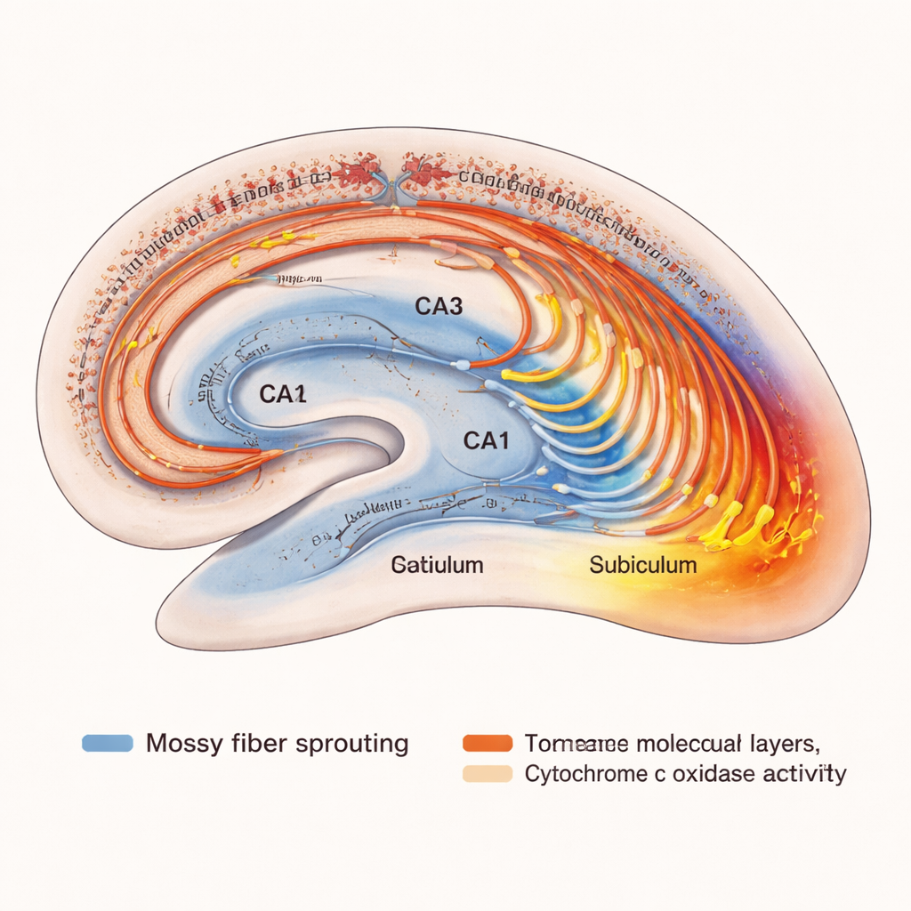
Energy Hotspots in a Hyperactive Network
Seizures are bursts of intense electrical activity that demand large amounts of energy. To gauge how local metabolism changes in MTLE, the authors stained for cytochrome c oxidase, a core enzyme in mitochondrial energy production. Compared with control brains, MTLE hippocampi showed reduced enzyme labeling in the already damaged CA1 area but increased labeling in the inner molecular layer and the subiculum. In other words, the very zones receiving the sprouted mossy fibers also appeared to be running “hot,” with a higher metabolic capacity. This pairing of structural rewiring and elevated energy use supports the idea that these regions may form a hyperactive hub that sustains or spreads seizures, even after much of the original circuitry has degenerated.
What This Means for People With Difficult-to-Treat Epilepsy
To a non-specialist, the message is that MTLE is not just a matter of dead tissue in the hippocampus; it is also a story of surviving nerve cells sending out new, possibly misguided, connections and of certain regions becoming metabolic hotspots. The study suggests that mossy fibers may form longer-than-normal routes from the dentate gyrus into CA1 and the subiculum, and that these rearranged pathways could help drive ongoing seizures in drug-resistant patients. By mapping both the new wiring and the energy landscape, the work hints at future strategies—from refined surgical targets to novel interventions aimed at these overactive pathways—that might one day control seizures while preserving as much healthy memory circuitry as possible.
Citation: Tu, T., Wan, L., Zhang, QL. et al. Putative long range mossy fiber sprouting and regional cytochrome c oxidase alteration in the hippocampus of patients with mesial temporal lobe epilepsy. Sci Rep 16, 5232 (2026). https://doi.org/10.1038/s41598-026-36148-3
Keywords: temporal lobe epilepsy, hippocampus, mossy fiber sprouting, neuronal loss, brain metabolism