Clear Sky Science · en
Alzheimer’s related dementia severity classification from magnetic resonance imaging using derivative-free optimization of convolutional neural network
Helping Doctors See Dementia Earlier
As populations age, many families worry about memory loss and dementia. Brain scans can reveal early changes linked to Alzheimer’s disease, but reading thousands of images by eye is slow and difficult, even for experts. This study presents a smart computer tool that looks at MRI brain scans and sorts people into four levels of dementia severity, from no dementia to moderate impairment. The system is designed to be both extremely accurate and light enough to run on ordinary hospital computers, making advanced image analysis more widely accessible.
A New Smart Helper for Brain Scans
The researchers focus on a type of artificial intelligence called a convolutional neural network, or CNN, which excels at spotting patterns in images. Instead of simply deciding whether someone has Alzheimer’s disease, their tool distinguishes four stages: non-dementia, very mild, mild, and moderate dementia. To do this, the team trained their model on large public collections of MRI scans that had already been labeled by experts. The goal was twofold: to reach near-perfect accuracy and to keep the model compact and fast, so that it could be practical for everyday clinical use rather than only for well-funded research labs.

Balancing the Data and Cleaning the Images
A key challenge in medical data is that not all stages of disease are represented equally. In these brain-scan collections, healthy and very mild cases are common, while moderate dementia scans are much rarer. Standard AI systems then tend to “play it safe” by overpredicting the common classes and missing early or moderate disease. To counter this, the authors used a two-step strategy: they first removed confusing borderline images and then created realistic synthetic examples of the underrepresented stages. Alongside this, they carefully prepared each MRI slice by filtering, isolating the brain from surrounding tissue, and normalizing brightness and contrast so that the model could focus on medically meaningful features such as shrinking of deep memory structures and widening of fluid-filled spaces.
Designing a Smaller, Smarter Network
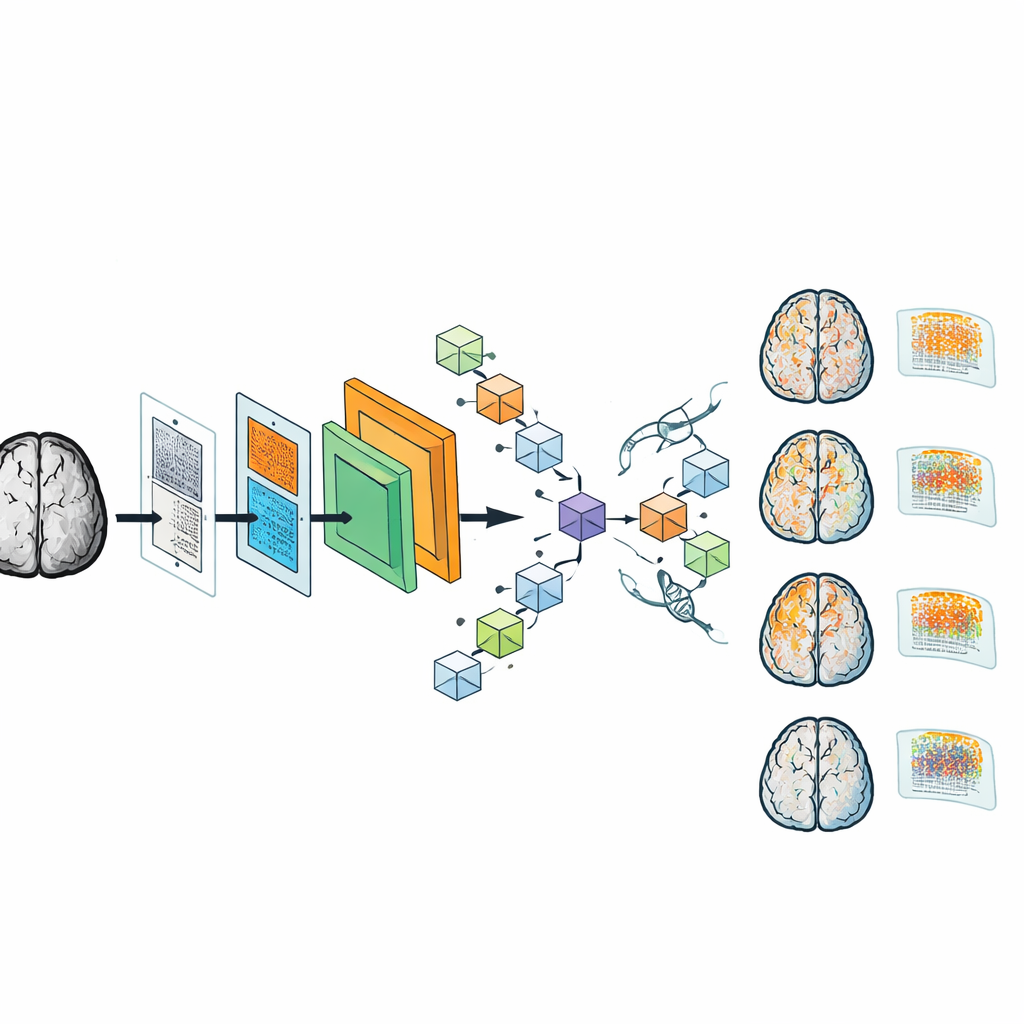
Instead of relying on traditional training methods that tweak parameters by following mathematical gradients, the team used a family of so-called derivative-free optimization techniques. They began with a larger, conventional CNN and then used evolutionary search and neural architecture search to evolve a simpler design with only three convolutional layers and far fewer filters. Bayesian optimization then fine-tuned how the network should be trained, while simulated annealing and pruning removed unnecessary connections after training. The end result, called DAPA-CNN, has about 85% fewer adjustable parameters than the starting model, uses around one quarter of the memory, and trains in less than two thirds of the time, yet still captures the crucial patterns in the scans.
Near-Perfect Performance and Clear Visual Explanations
Despite its smaller size, DAPA-CNN proved remarkably accurate. On a major Alzheimer’s dataset, it correctly assigned dementia stage in almost every case, with accuracy and other performance measures all hovering around 99%. The model also generalized well to a separate multi-center dataset collected on different scanners, suggesting that it is robust to real-world variations in imaging. To make the system more transparent for clinicians, the authors generated class activation maps—heat-like overlays that show which regions of the brain influenced a given decision. In early stages, these maps emphasize areas involved in memory that are known to deteriorate first in Alzheimer’s disease; in later stages, they spread to wider cortical regions, aligning with clinical understanding of disease progression.
What This Means for Patients and Clinics
For non-specialists, the main message is that the study offers a fast, compact, and interpretable tool to grade dementia severity from routine MRI scans. By balancing the training data and carefully shaping the network with derivative-free optimization, the authors created a model that can run on modest hardware while delivering close to perfect accuracy and highlighting the brain regions behind its decisions. If confirmed in future prospective clinical trials, such technology could support earlier diagnosis, more consistent staging across hospitals, and better tracking of how Alzheimer’s disease changes the brain over time.
Citation: Ganesan, S.K., Velusamy, P., Parthsarathy, P. et al. Alzheimer’s related dementia severity classification from magnetic resonance imaging using derivative-free optimization of convolutional neural network. Sci Rep 16, 10077 (2026). https://doi.org/10.1038/s41598-026-36037-9
Keywords: Alzheimer’s disease, brain MRI, dementia staging, deep learning, medical imaging AI