Clear Sky Science · en
Perivascular pathology, not macrovascular complexity, governs glymphatic-related dysfunction in preclinical cerebral small vessel disease
Why Brain Drainage Matters Before Symptoms Appear
Long before a stroke or memory loss shows up, tiny blood vessels deep inside the brain can start to fail silently. This study looks at how the brain’s waste‑clearance system, sometimes called the “glymphatic” pathway, is affected in seemingly healthy, working‑age adults. By pairing advanced brain scans with measures of vessel shape and microscopic fluid flow, the authors ask a deceptively simple question: is early trouble driven more by the big arteries supplying the brain, or by damage around the smallest vessels buried within it?
Tiny Fluid Channels That Keep the Brain Clean
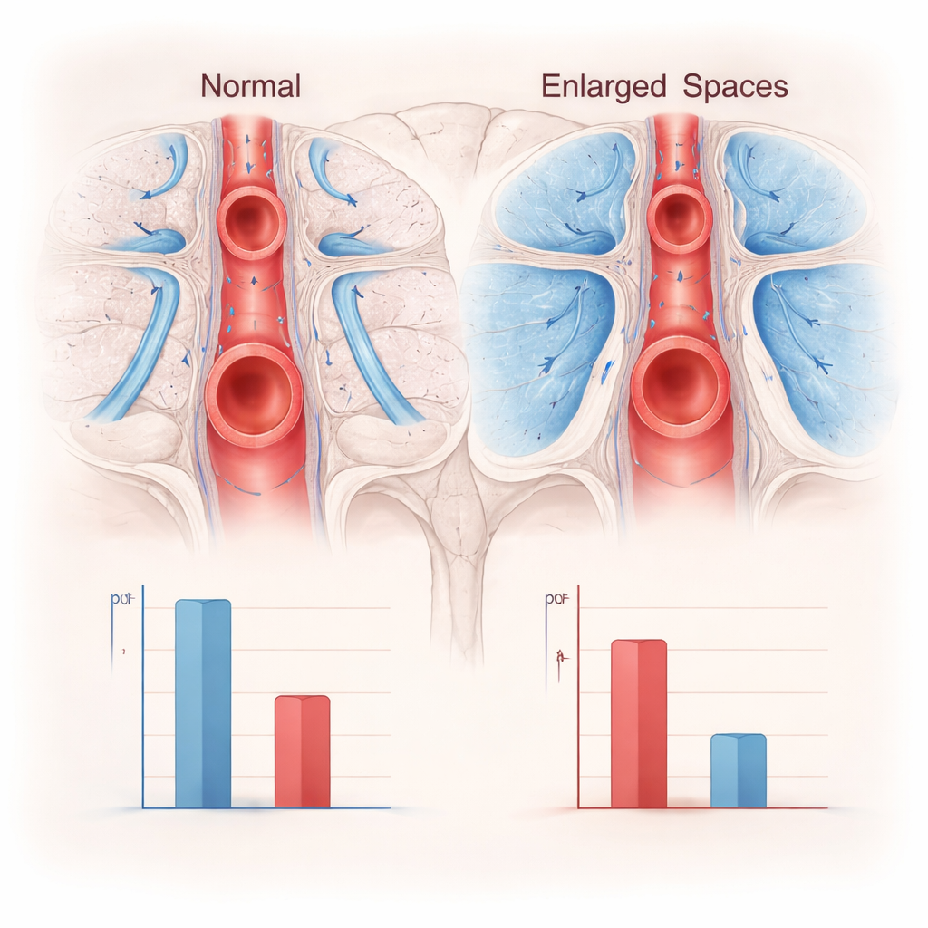
The brain constantly produces waste products as nerve cells work. To stay healthy, it relies on fluid that moves along the outsides of blood vessels, washing away debris through narrow corridors called perivascular spaces. When these channels swell and become visibly enlarged on MRI, they are thought to signal that this cleaning system is struggling. These enlarged perivascular spaces, or ePVS, are also a hallmark of cerebral small vessel disease, a slow, often “silent” process that can eventually lead to stroke and dementia. The researchers focused on people who had no symptoms and only low‑to‑moderate cardiovascular risk, to capture this process at a very early, preclinical stage.
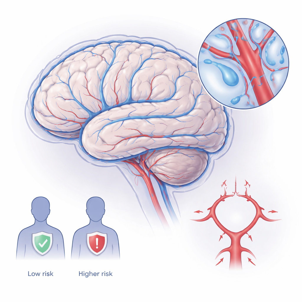
Testing Big Arteries Versus Small Vessel Damage
To see whether the shape of large brain arteries matters for this cleaning system, the team examined the circle of Willis – a ring‑like junction of major arteries at the base of the brain. Using a mathematical measure called fractal dimension, they quantified how intricate and space‑filling this arterial network was, a proxy for how well it might distribute blood and pulsations that drive fluid movement. At the same time, they used a diffusion MRI method (the DTI‑ALPS index) that captures how easily water moves along perivascular pathways, serving as an indirect readout of glymphatic‑related activity. Finally, they graded each person’s ePVS burden on structural MRI and measured standard thinking skills, including working memory and processing speed.
What the Brain Scans Revealed
Among 60 adults, about 4 in 10 already showed visible ePVS, even though none had diagnosed brain disease and overall cardiovascular risk scores were low. People with ePVS tended to be slightly older, more likely to be on blood pressure medication, and had higher long‑term vascular risk scores, fitting with the idea that these tiny lesions reflect cumulative vessel stress. Crucially, the ePVS group had both simpler‑looking circles of Willis and markedly lower DTI‑ALPS values, indicating reduced fluid movement along perivascular routes. At first glance, the complexity of the large arteries did relate to the fluid‑flow measure, and both were tied to ePVS burden.
Microvessels Take Center Stage
However, once the researchers adjusted for age, sex, blood pressure treatment, overall vascular risk, and ePVS, the apparent link between big‑artery complexity and fluid‑flow largely disappeared. Statistical models showed that the single strongest predictor of reduced perivascular diffusivity was the presence of ePVS themselves. In other words, how damaged or swollen the small‑vessel surroundings were mattered far more than how elaborate the main arterial ring looked. A more detailed mediation analysis confirmed that changes in the circle of Willis did not explain how ePVS were linked to poorer fluid dynamics. Cognitive test scores were generally normal, with only subtle, non‑significant trends suggesting that individuals with better working memory and processing speed tended to have more efficient perivascular diffusion.
What This Means for Protecting Brain Health
For a non‑specialist, the message is that early brain “plumbing” problems show up first around the tiniest vessels, not in the grand architecture of the main arteries. Visible enlarged perivascular spaces on MRI stand out as a practical, clinically relevant marker that the brain’s waste‑clearance system is under strain, even in people who feel well and perform normally on standard thinking tests. By contrast, the fine‑grained geometry of the circle of Willis, while interesting and altered in those with small‑vessel damage, did not independently govern this clearance measure. These findings support a shift toward monitoring microvascular health as a way to spot and potentially prevent small vessel disease and related cognitive decline long before symptoms emerge.
Citation: Hein, Z.M., Che Mohd Nassir, C.M.N. Perivascular pathology, not macrovascular complexity, governs glymphatic-related dysfunction in preclinical cerebral small vessel disease. Sci Rep 16, 4528 (2026). https://doi.org/10.1038/s41598-026-36001-7
Keywords: brain waste clearance, small vessel disease, perivascular spaces, glymphatic system, brain MRI