Clear Sky Science · en
Multiparametric MRI radiomics nomogram predicts synchronous distant metastasis in rectal cancer
Why predicting cancer spread matters
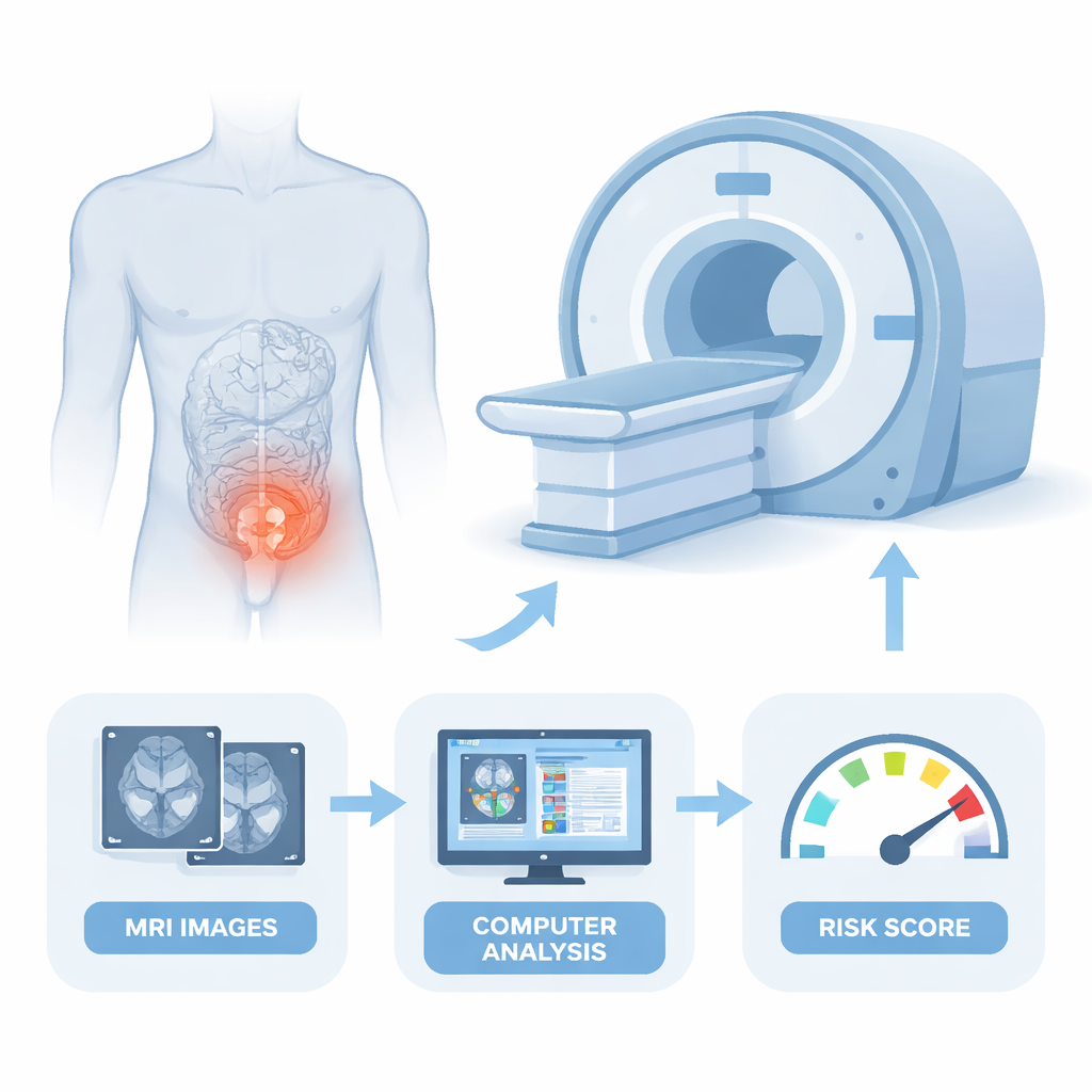
When rectal cancer is first diagnosed, doctors urgently need to know whether the disease has already begun to spread to distant organs such as the liver or lungs. Catching this spread early can open the door to curative surgery and more tailored treatment, while missing it can mean unnecessary operations or delayed care. This study explores whether advanced computer analysis of routine MRI scans can flag patients at high risk of hidden spread right from the start.
Looking for clues hidden in scans
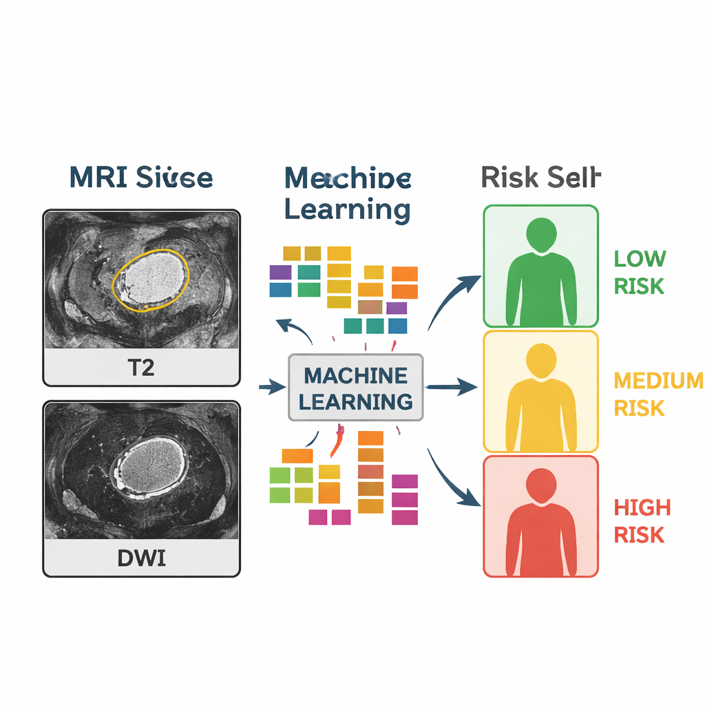
Modern MRI machines do more than create sharp pictures; they also capture subtle patterns of brightness and texture inside tumors that the human eye cannot easily see. The researchers used a technique called “radiomics,” which turns these patterns into thousands of numerical features. They focused on two common MRI types used for rectal cancer: T2-weighted images, which show anatomy in fine detail, and diffusion-weighted images, which highlight how water moves through tissues and can reflect how densely packed cancer cells are.
Building a risk score from images and blood tests
The team reviewed data from 169 people with rectal cancer who had MRI and CT scans before any major treatment. Nearly half already had distant metastases at diagnosis. From more than 1,600 image-based features, they used statistical filters and machine-learning methods to whittle the list down to a small set that best separated patients with and without distant spread. They then combined these selected image features with simple clinical information, including age, tumor stage on MRI, and two common blood markers (CEA and CA19-9), to create a single prediction tool known as a nomogram—a kind of visual risk calculator.
How well did the tool work?
To test their approach, the researchers split the patients into a larger training group and a smaller test group. Models that used either clinical data alone or radiomics alone already showed reasonable ability to distinguish patients with distant spread from those without. However, when they blended the two—mixing image-derived features with clinical factors—the performance improved noticeably. In the independent test group, the combined nomogram correctly separated patients with and without synchronous distant metastasis about nine times out of ten, with a good balance between catching high-risk patients and avoiding false alarms. Additional checks suggested that the tool’s predictions lined up closely with what actually happened and that using it could provide more clinical benefit than relying on standard measures alone.
What the images revealed about aggressive tumors
The computer analysis highlighted that texture details from diffusion-weighted MRI were especially informative. Tumors whose diffusion images showed greater internal irregularity—an imaging sign of tissue chaos and mixed cell density—were more likely to be linked with distant spread. In other words, the more uneven and complex the tumor looked at the microscopic level, as captured indirectly by the scan, the higher the likelihood that cancer cells had already escaped to other parts of the body. This supports the idea that advanced imaging can serve as a non-invasive window into tumor behavior, not just its size and shape.
What this could mean for patients
For people newly diagnosed with rectal cancer, a tool like this MRI-based nomogram could help doctors quickly sort patients into lower- and higher-risk groups for distant metastasis before surgery or major therapy begins. Those flagged as high risk might be sent for more intensive whole-body imaging, closer follow-up, or more aggressive treatment plans, while lower-risk patients could be spared unnecessary tests and anxiety. Although the study was done at a single center and still needs confirmation in larger, multi-hospital trials, it points toward a future where routine scans and simple blood tests are combined with machine learning to guide more personalized, timely care.
Citation: Jiang, H., Guo, W., Lin, X. et al. Multiparametric MRI radiomics nomogram predicts synchronous distant metastasis in rectal cancer. Sci Rep 16, 5759 (2026). https://doi.org/10.1038/s41598-026-35973-w
Keywords: rectal cancer, MRI radiomics, metastasis risk, machine learning in medicine, cancer imaging