Clear Sky Science · en
Deep learning-based classification of thyroid nodules using uncertainty-aware multi-modal ultrasound imaging
Why thyroid lumps matter to everyone
Small lumps in the thyroid gland are extremely common, especially as people age. Most are harmless, but a minority are cancerous and need timely treatment. Today, doctors mainly rely on ultrasound scans and needle biopsies to tell the difference. Ultrasound is safe and widely available, yet its interpretation can vary from one examiner to another, leading some people to undergo unnecessary invasive tests while others may be missed. This study explores how combining multiple types of ultrasound with an artificial intelligence system can sharpen thyroid cancer diagnosis and even tell doctors how confident the computer is in its answer.
Looking at the same nodule in several ways
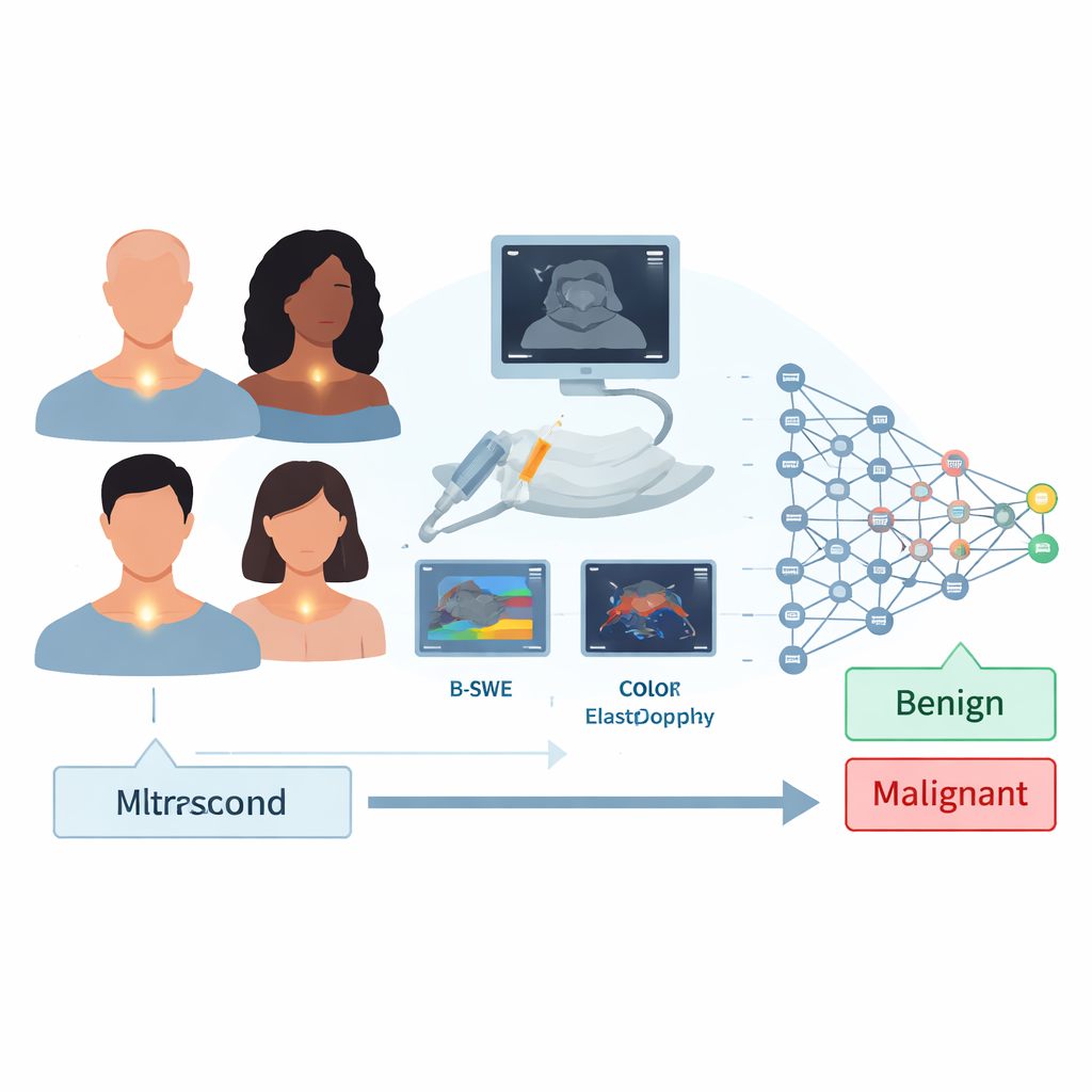
Ultrasound is not just a single kind of picture. Standard “B-mode” ultrasound shows the shape and texture of a thyroid nodule. Shear wave elastography adds information about how stiff the tissue is, which often differs between benign and malignant growths. Color Doppler highlights blood flow patterns inside and around the nodule. Previous research usually focused on only one of these views, or simple combinations, and did not clearly address how reliable each source of information was when feeding it into a computer model.
Building a smarter, leaner AI for thyroid scans
The researchers prospectively collected images from 506 thyroid nodules in 422 patients who were already scheduled for biopsy at a single medical center. For each nodule, they acquired B-mode, shear wave elastography, and color Doppler images from different machines within the same ultrasound family. They then designed a custom deep learning network that first used a compact, pretrained image-recognition backbone and added a lightweight “head” tailored for medical ultrasound. This head used special layers that mix different kinds of feature extraction and attention, helping the model focus on the most informative regions of each image while keeping the overall architecture relatively small and efficient.
Letting the computer admit when it is unsure
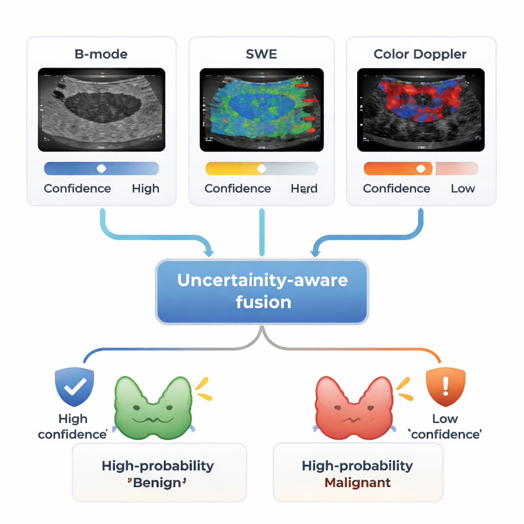
A key innovation of the study is an uncertainty-aware fusion strategy. Instead of simply averaging predictions from the three ultrasound types, the system estimates how confident each branch is for a given patient. It does this by running the model multiple times with small internal variations and measuring how stable the predictions are. If one modality, such as color Doppler, gives a shaky or inconsistent answer, its influence on the final decision is reduced or even set to zero. By contrast, a modality that is both accurate and confident, such as the combination of B-mode and shear wave elastography in many cases, is given more weight. This mirrors how human radiologists naturally trust clear, high-quality images more than noisy or ambiguous ones.
How well did the system work?
Using a rigorous cross-validation design, the combined three‑modalities system correctly classified thyroid nodules with an accuracy of about 95 percent and an area under the ROC curve of 0.97. Sensitivity—the ability to detect cancers—was especially high at 98 percent, while specificity for ruling out cancer reached 92 percent. Single imaging types and two‑way combinations performed less well, showing that multi‑modal input truly added value. The uncertainty‑aware fusion method also outperformed simpler ways of combining predictions, particularly in handling conflicting or unreliable inputs. In comparisons with many well‑known deep learning models adapted to the same data, the custom architecture matched or exceeded their performance despite using fewer layers and being more compact.
What this means for patients and doctors
For patients with thyroid nodules, this work points toward a future in which a routine ultrasound exam could provide not just a yes‑or‑no risk estimate for cancer, but also a sense of how trustworthy that estimate is. A highly confident benign prediction might help avoid unnecessary biopsies, while a high‑uncertainty result could trigger extra imaging, a second opinion, or closer follow‑up. Although the study was conducted at a single center and still needs confirmation across different hospitals and ultrasound machines, the results suggest that combining several ultrasound views with an uncertainty‑aware AI system can make thyroid cancer diagnosis both more accurate and more transparent, potentially improving care while reducing needless procedures.
Citation: Saini, M., Parvar, T.A., Velarde, M. et al. Deep learning-based classification of thyroid nodules using uncertainty-aware multi-modal ultrasound imaging. Sci Rep 16, 4938 (2026). https://doi.org/10.1038/s41598-026-35965-w
Keywords: thyroid nodules, ultrasound imaging, deep learning, cancer diagnosis, medical AI