Clear Sky Science · en
A new combined reduction anatomical plate for the treatment of acetabular anterior column and posterior hemi-transverse fractures: a finite element analysis study
Why broken hips are so hard to fix
When the socket of the hip joint shatters in a car crash or fall, surgeons face a delicate puzzle deep inside the pelvis. These acetabular fractures must be repaired precisely to let people walk without pain or early arthritis. Yet current metal plates used to hold the broken bone together can be difficult to shape, may not fit well, and sometimes fail, leading to more surgery. This study introduces a newly designed plate system and uses computer modeling to test whether it can stabilize these complex fractures more safely and reliably.
A hidden break in the hip socket
The work focuses on a particularly tricky injury called an anterior column–posterior hemi-transverse fracture of the acetabulum. In this pattern, the front and part of the back of the hip socket crack, often driving the ball of the thigh bone inward. The break frequently extends into a thin bony area on the inside of the pelvis known as the quadrilateral plate, which is hard for surgeons to see and reach. Older patients, whose bones are more fragile, are especially vulnerable. If the socket is not restored to a smooth, round shape, the blood supply to the hip can suffer and the joint can quickly wear out, causing permanent disability.
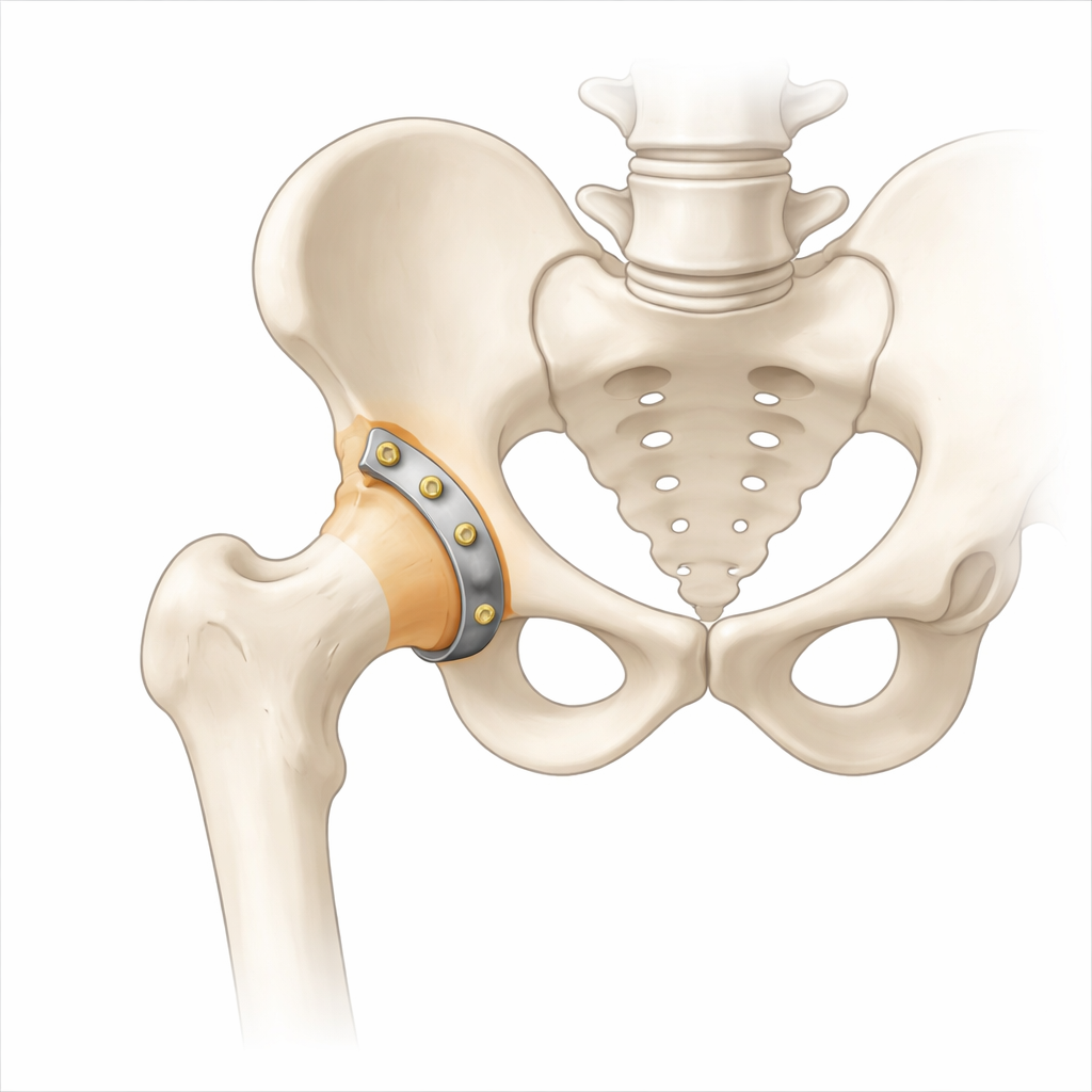
A plate shaped to fit the pelvis
To address these challenges, the research team developed the Combined Reduction Anatomical Plate, or CORAP. Instead of a single flat strip of metal, CORAP is a two-part system: a locking plate that curves along the back of the pelvis above a natural notch in the bone, and a reduction plate that hugs the inner surface around the hip socket and quadrilateral plate. Both parts are pre-shaped using detailed measurements of pelvic anatomy, so the plate is meant to match the bone closely without extensive bending in the operating room. The device is intended to give surgeons more secure screw positions while reducing the risk that screws stray into the hip joint.
Testing plates inside a virtual pelvis
Because it is difficult to compare many metal designs in real patients, the authors turned to the finite element method, a type of computer simulation widely used in engineering. They created a three-dimensional model of a human hemipelvis from CT scans of a healthy young volunteer, then digitally cut the bone to reproduce the target fracture pattern. Onto this virtual broken pelvis they mounted four different fixation systems: the new CORAP, a pair of traditional plates spanning both columns of the socket, a plate designed to support the inner quadrilateral surface, and a combination of plates along the front and inner back of the pelvis.
The model included key ligaments and joint cartilage, and the researchers simulated three everyday body positions: standing, sitting, and lying on the injured side. For each posture they applied loads of 200, 400, and 600 newtons—roughly representing partial to near-full weight bearing—and calculated how stress spread through the plates and screws, how much the pelvis as a whole moved, and how much the fracture edges rubbed or separated.
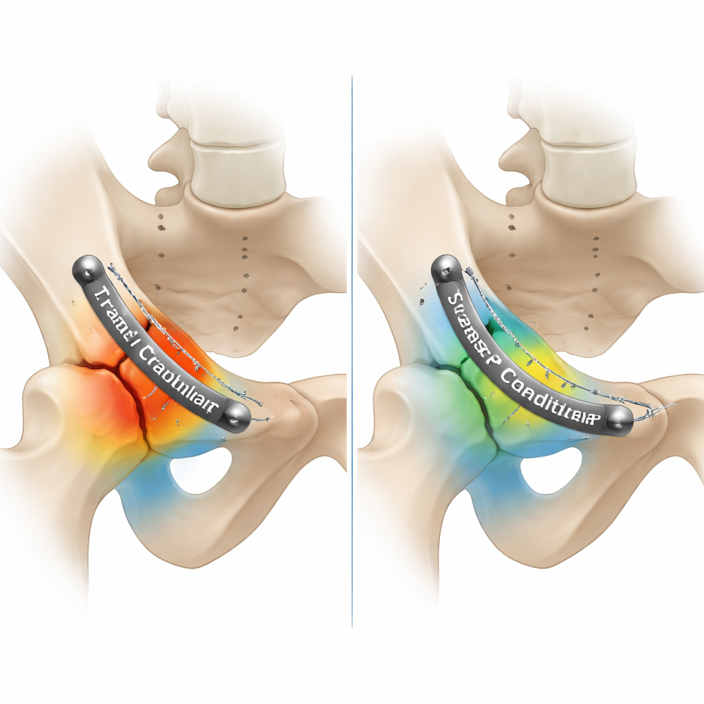
How the new design spreads the load
The simulations showed that, under all tested positions and forces, CORAP kept stresses in the metal well below the strength limit of the titanium alloy. Compared with two of the traditional single-plate systems, CORAP distributed stress more evenly and avoided pronounced hotspots where a plate or screw might crack. Its stiffness—how much it resisted bending—was slightly lower than that of the double-column plate construct, which unsurprisingly had the smallest overall motion because it used two robust plates. Yet CORAP allowed only tiny movements at the fracture line, on the order of a few thousandths of a millimeter, comparable to the other methods and within the range thought to encourage the formation of healing bone callus.
What this could mean for patients
From a patient’s perspective, the most important question is whether a new device keeps the hip stable enough to heal while limiting surgical trauma. This study suggests that CORAP offers a good compromise: it behaves almost as stably as the more invasive double-plate technique, but is simpler and better matched to the natural curves of the pelvis. The computer models indicate that even when a person with this implant stands, sits, or lies on the injured side, the plate and screws are unlikely to fail, and the small, controlled motion between bone fragments should favor solid healing. Although further testing in cadavers and clinical trials is still needed, the findings support CORAP as a promising, safe option for repairing difficult hip socket fractures.
Citation: Chongshuai, B., Jun, A. & Lin, C. A new combined reduction anatomical plate for the treatment of acetabular anterior column and posterior hemi-transverse fractures: a finite element analysis study. Sci Rep 16, 5306 (2026). https://doi.org/10.1038/s41598-026-35856-0
Keywords: hip fracture, acetabulum, internal fixation, biomechanics, finite element analysis