Clear Sky Science · en
Association of choroid plexus volume with brain atrophy and glucose metabolism in multiple system atrophy
Why brain fluid factories matter in a rare movement disease
Deep inside the brain sits a little-known tissue called the choroid plexus, which constantly produces the fluid that bathes and cleans our nervous system. This study asks a deceptively simple question with big implications: in multiple system atrophy (MSA)—a fast-moving, Parkinson’s-like disorder—does this fluid factory change in size, and do those changes mirror the brain damage seen on scans? The answers could point to a new, easily measured marker of disease burden and shed light on how the brain’s cleaning system fails in neurodegeneration.
The brain’s hidden filter and cleaning crew
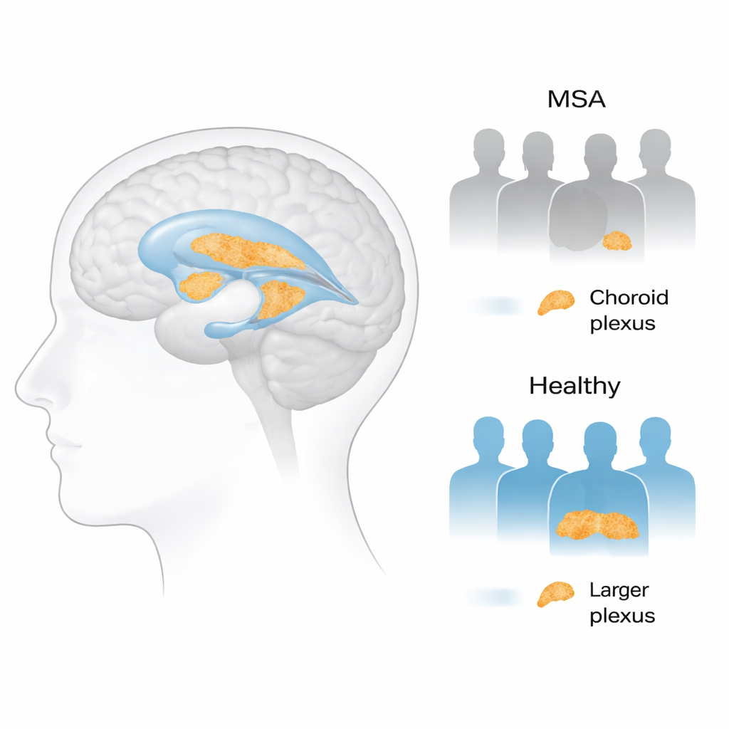
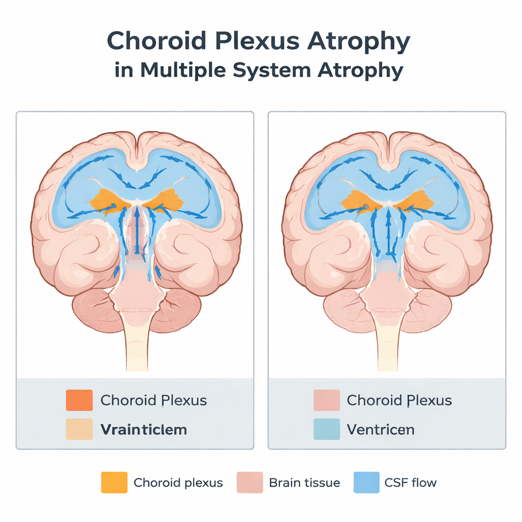
The choroid plexus sits inside fluid-filled spaces called ventricles and pumps out hundreds of milliliters of cerebrospinal fluid each day. That fluid cushions the brain, delivers nutrients and hormones, and helps wash away toxic waste through a recently described “glymphatic” clearance system. Earlier work in Alzheimer’s, Parkinson’s disease, and other brain disorders found that this tissue tends to grow larger, possibly as a reaction to increasing waste and inflammation. But almost nothing was known about how it behaves in MSA, a rare condition marked by stiffness, problems with balance and coordination, and failure of automatic functions like blood pressure control.
Comparing patients and healthy volunteers
The researchers examined 87 people with MSA and 84 healthy older adults. All participants had detailed brain MRI scans to measure the volume of the choroid plexus and other brain structures, and most also underwent PET scans that show how much sugar (glucose) different brain areas are using—a proxy for how active and healthy those regions are. The team also recorded clinical ratings of movement and disability, and sorted patients into two main clinical forms: a parkinsonian type that looks more like classic Parkinson’s disease, and a cerebellar type that mainly affects balance and coordination.
A surprising shrinkage instead of swelling
Contrary to patterns seen in Alzheimer’s and Parkinson’s disease, people with MSA had a markedly smaller choroid plexus than healthy volunteers, even after accounting for age and sex. Both MSA subtypes showed this shrinkage to a similar degree. Patients also had reduced volume in key structures such as the hippocampus and greater damage in deep white matter, signs of widespread brain injury. Within the MSA group, those with smaller choroid plexus volume tended to have more pronounced loss of tissue in the cerebellum and certain deep brain regions involved in movement and body regulation. However, choroid plexus size did not track directly with bedside movement scores, suggesting that standard clinical scales may not be sensitive to this aspect of disease biology.
Linking brain cleaning power to energy use
When the team looked at PET scans, they found that larger choroid plexus volume was associated with higher glucose metabolism—essentially stronger energy use—in the brainstem, cerebellar white matter, and parts of the thalamus and nearby regions, all areas known to be vulnerable in MSA. Patients with more shrunken choroid plexus tissue showed lower activity in these regions, consistent with more advanced neurodegeneration. Importantly, similar relationships were not seen in healthy volunteers, hinting that this coupling between the choroid plexus and brain metabolism is specific to the disease state rather than a general feature of normal aging.
What this could mean for patients
Taken together, the findings suggest that in MSA the brain’s fluid-producing tissue withers rather than enlarges, and that this loss goes hand in hand with structural damage and reduced energy use in especially vulnerable brain regions. One possibility is that as the choroid plexus shrinks, it produces less fluid, further weakening the brain’s ability to flush out toxic proteins and maintain stability—a vicious cycle that accelerates injury in movement and balance centers. Although more work, especially long-term follow-up studies, is needed, choroid plexus volume measured on routine MRI could become a practical imaging marker to gauge overall disease burden and to test whether future treatments help preserve the brain’s internal cleaning system in multiple system atrophy.
Citation: Park, C.J., Sun, Y., Jeong, HJ. et al. Association of choroid plexus volume with brain atrophy and glucose metabolism in multiple system atrophy. Sci Rep 16, 5551 (2026). https://doi.org/10.1038/s41598-026-35850-6
Keywords: multiple system atrophy, choroid plexus, brain atrophy, glucose metabolism, neurodegeneration