Clear Sky Science · en
High-accuracy brain tumor detection method based on deep learning
Why spotting brain tumors sooner matters
Brain tumors are among the deadliest diseases of the nervous system, and catching them early can make the difference between life and death. Today, doctors usually search for tumors by carefully inspecting magnetic resonance imaging (MRI) scans by eye—a demanding task that can be slow, subjective, and easy to misjudge when the tumor is tiny or its edges are blurry. This study describes a new artificial intelligence (AI) system that aims to help radiologists find three common types of brain tumors more quickly and accurately, potentially improving treatment planning and patient outcomes.
A smarter digital assistant for MRI scans
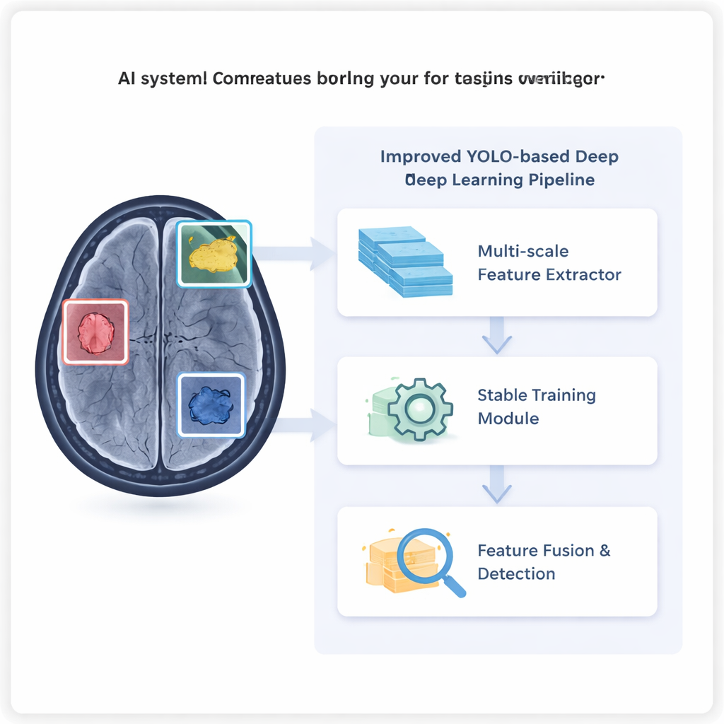
The researchers build on a popular real-time object detection family of algorithms known as YOLO, widely used to locate objects in everyday photos and videos. Instead of cars or pedestrians, however, this upgraded version is trained to find meningiomas, pituitary tumors, and gliomas in brain MRI images. Using a public dataset from the Kaggle platform and additional CT scans from Radiopaedia, the team trained their system to draw boxes around tumors and label their type. They then compared its performance with several state-of-the-art AI models to see whether the new design really helps doctors see more of what matters and less of what does not.
Seeing the small and subtle signs
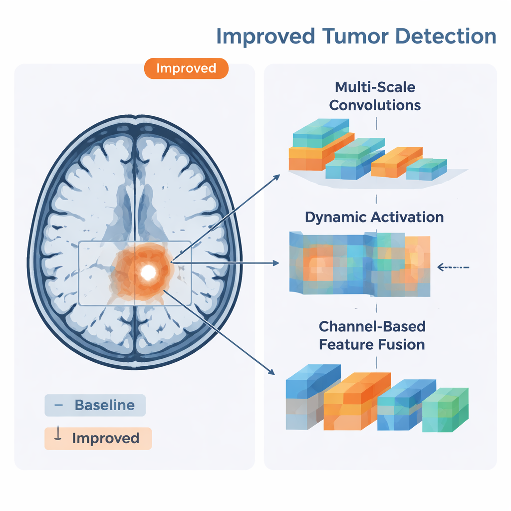
A key challenge in brain imaging is that tumors vary widely in size and shape, and some blend almost seamlessly into surrounding tissue. To address this, the authors introduced a new component they call the A2C2f-Mona module. In simple terms, it looks at each scan through several "lenses" of different sizes at once, capturing both fine details and broader patterns. This multi-scale view helps the system pick up subtle changes in texture and intensity that might mark the border of a tumor. In tests, this design particularly improved detection of small or faint lesions, where standard models often hesitate or miss the target entirely.
Keeping learning stable and focused
Training deep neural networks often relies on mathematical tricks called normalization layers to keep internal signals from blowing up or fading away. But in medical imaging, where batches of images can be small and varied, these tricks can become unstable and computationally heavy. The study replaces them with a lighter "dynamic" transformation, dubbed C2PSA-DyT, which uses a smooth mathematical curve to keep activations in a reasonable range without the usual overhead. This change makes the model more stable to train and frees up capacity for other improvements, helping it maintain consistent performance across many different scans.
Combining clues from different depths
Another hurdle is how to merge coarse, high-level information (such as where a suspicious region lies) with crisp, low-level details (like exact edges and textures). The authors tackle this with a CGAFusion module, which works a bit like a spotlight that brightens the most informative channels of the image while dimming less useful ones. By blending shallow and deep features with learned attention weights, the system becomes better at outlining tumors whose margins fade into normal tissue and at distinguishing tumors from look-alike structures such as blood vessels or the lining of the brain. Visual explanations using Grad-CAM heatmaps show that the model’s attention tends to fall squarely on the true tumor regions, aligning well with expert judgment.
What the results mean for patients and doctors
On the brain tumor test set, the new system achieved a precision of about 94% and a recall of 88%, both higher than the strongest YOLO baseline and several other leading detectors. It was especially good at finding pituitary tumors, a category where missed cases can have serious hormonal and vision consequences, and it modestly but significantly improved detection of hard-to-spot gliomas. Crucially, the method still runs fast enough for real-time use, suggesting it could be integrated into hospital imaging workflows as a second pair of eyes for radiologists. While the authors note that larger, multi-center studies and true 3D imaging will be needed before clinical deployment, their work shows that carefully designed AI can make brain tumor detection both more accurate and more reliable—helping doctors focus on complex decisions while the algorithm tirelessly scans every pixel.
Citation: Ye, W., Chen, Z., Sun, X. et al. High-accuracy brain tumor detection method based on deep learning. Sci Rep 16, 5122 (2026). https://doi.org/10.1038/s41598-026-35783-0
Keywords: brain tumor detection, MRI imaging, deep learning, object detection, medical AI