Clear Sky Science · en
A computational intelligence approach for classifying dental caries in X-ray images using integrated fuzzy C-means clustering with feature reduction and a weighted matrix scheme
Why smarter cavity detection matters
Dentists rely heavily on X-ray images to spot tooth decay that isn’t visible to the naked eye. But these images are often noisy, blurry, and low in contrast, making early cavities easy to miss. This study introduces a new computer-based method that helps identify decayed areas in dental X-rays more accurately and consistently. By doing so, it could support dentists in catching problems earlier, planning better treatments, and improving access to high-quality care in clinics that lack cutting-edge equipment.
The challenge of reading dental X-rays
Tooth decay, or dental caries, affects people of all ages and can lead to pain, infection, and tooth loss if not treated early. Traditional X-rays show a flat, two-dimensional snapshot of complex three-dimensional structures. Small lesions can be hidden by overlapping tissues, blurred by patient movement, or obscured by metal fillings. On top of that, many hospitals—especially in resource-limited regions—still rely on basic X-ray machines that produce images with uneven brightness and considerable noise. These factors make it hard, even for experienced dentists, to reliably distinguish a tiny patch of early decay from normal variations in tooth structure.
Limitations of current AI approaches
In recent years, researchers have turned to artificial intelligence to read dental images. Deep learning systems, in particular, can perform very well, but they come with major drawbacks. They typically need thousands of carefully labeled images, which must be annotated by dental experts—a slow and expensive process. They also require powerful computers and graphical processors that many hospitals do not have. Even when such systems work well, they often act as “black boxes,” providing little insight into why a particular region was labeled as decayed or healthy. Existing methods also struggle with subtle, early-stage lesions and can be sensitive to differences in scanners, image quality, and patient populations.
A new way to let the data speak
This study proposes a different strategy based on an enhanced form of fuzzy C-means clustering, a technique that groups pixels in an image by similarity. Instead of assuming that all image features are equally important, the new method—called FCM-FRWS—automatically learns which characteristics matter most for separating decay from healthy tissue. It assigns a weight to each feature (such as local brightness, texture, or position), gradually downplaying those that add confusion and emphasizing those that clearly mark caries. Features that consistently contribute little are removed altogether, reducing noise and speeding up the process. This clustering is combined with smart image preparation steps: first, X-rays are normalized to a common contrast level, then smoothed to reduce random noise, and finally cleaned using simple shape-based operations so that the tooth outlines and potential cavities are easier to follow. 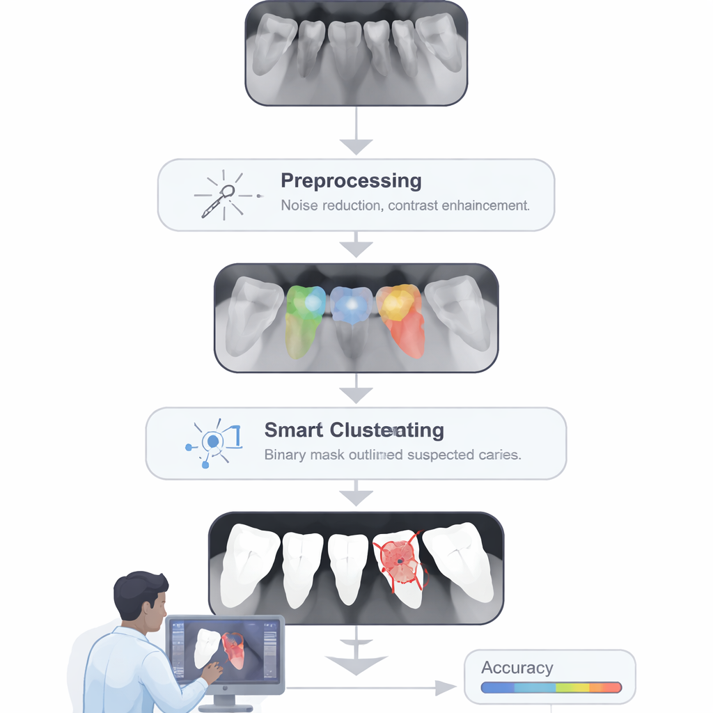
Sharpening the view of decay
After the weighted clustering has roughly separated likely tooth, background, and suspicious regions, the method applies a classic but powerful tool called Otsu’s thresholding to split pixels more cleanly into “lesion” and “non-lesion” groups based on their brightness. A morphological dilation step then slightly expands and connects fragmented spots so that each patch of decay is represented as one coherent region, not scattered dots. The complete pipeline—preprocessing, feature-weighted clustering, and refined thresholding—was tested on 890 X-ray images from hospitals in Northeastern Thailand, including both adults and children. Ground-truth markings from five experienced dentists were used as a reference. On average, the system correctly classified more than 91% of pixels, with similar high scores for sensitivity (detecting true decay), specificity (avoiding false alarms), and a strong overlap with dentists’ markings. Internal tests across different subsets of the data showed the method remained stable and did not simply memorize examples.
How this could help patients and clinics
Unlike many modern AI tools, this approach does not require a large labeled training set or specialized hardware, and it runs efficiently on an ordinary computer. That makes it attractive for smaller hospitals, teaching clinics, and practices in low-resource settings that still depend on standard X-ray machines. The method can act as a second reader, flagging suspicious areas for the dentist to review, especially in early, hard-to-see stages of decay. While it is not a replacement for clinical judgment and still has limits in very noisy or complex cases, the study shows that carefully designed, transparent algorithms can significantly improve cavity detection without the computing demands of deep learning. In the long run, such tools could be integrated directly into X-ray viewing software, quietly working in the background to help ensure that fewer cavities go unnoticed. 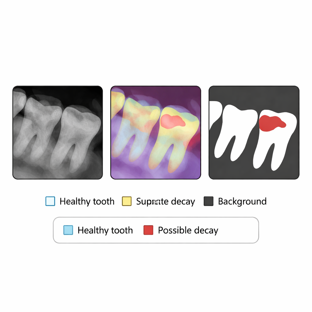
Citation: Wisaeng, K., Muangmeesri, B. A computational intelligence approach for classifying dental caries in X-ray images using integrated fuzzy C-means clustering with feature reduction and a weighted matrix scheme. Sci Rep 16, 5000 (2026). https://doi.org/10.1038/s41598-026-35735-8
Keywords: dental caries, X-ray imaging, medical image segmentation, fuzzy clustering, computer-aided diagnosis