Clear Sky Science · en
A validation of 3D imaging for non-invasive, tech-assisted diagnosis of caries and erosive tooth wear in primary teeth – an in vitro study
Why Your Child’s Tooth Scan Matters
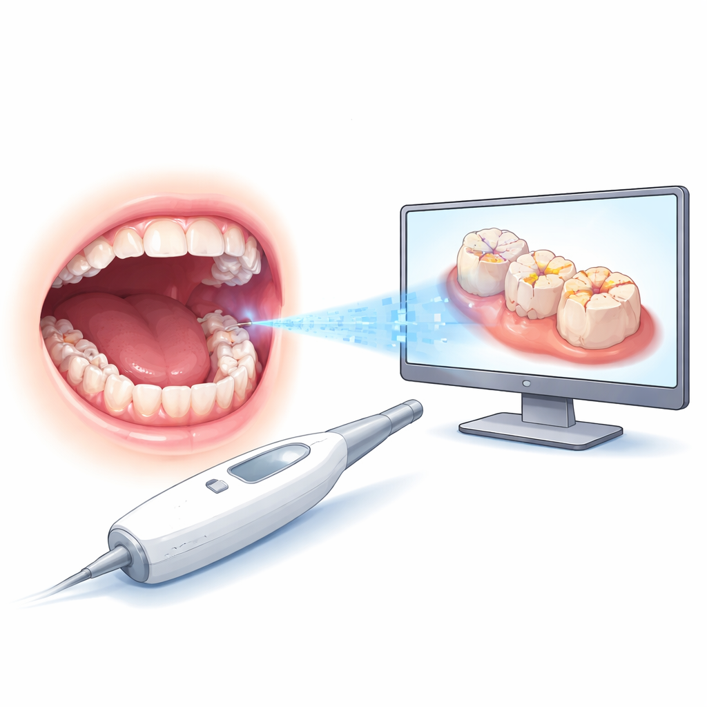
Parents today are used to digital tools in medicine, from phone apps to virtual visits. Dentistry is no exception: new 3D cameras can scan a child’s mouth without X-rays or poking instruments. This study asked a simple but important question: can colorful 3D scans of baby teeth spot cavities and early enamel wear about as well as a dentist looking directly in the mouth? The answer, based on hundreds of extracted primary teeth, is largely yes—especially for more advanced problems—suggesting these scans could soon become a routine helper in children’s dental care.
Everyday Problems in Tiny Teeth
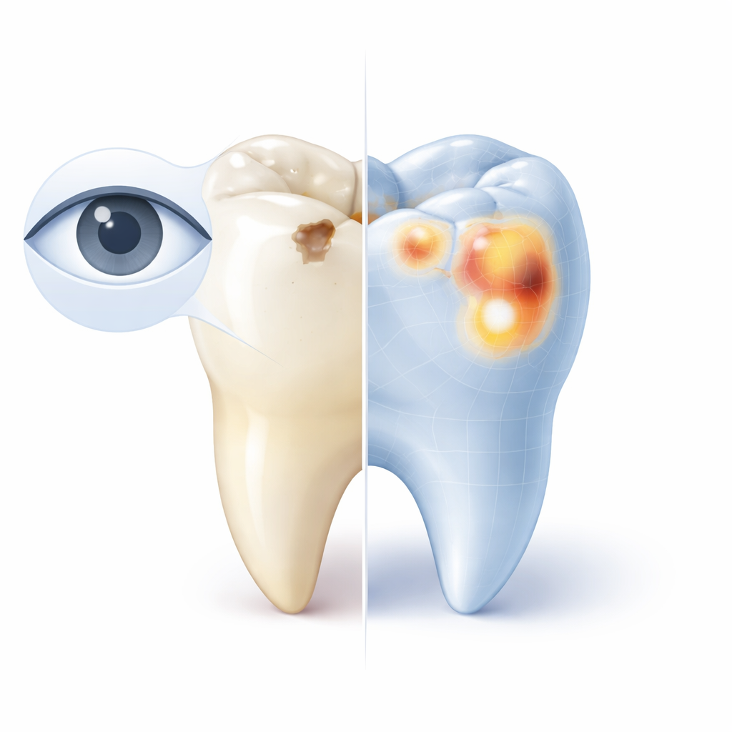
Tooth decay and erosion are among the most common health issues in children. Baby teeth have thinner, softer enamel than adult teeth, so damage can develop and spread quickly. Dentists usually rely on what they see with the naked eye, sometimes guided by standard scoring systems that rate how deep a cavity is or how much enamel has been worn away. But this visual judgment can vary from one clinician to another, and early, subtle changes are easy to miss. Catching these early spots is crucial, because they can often be reversed or slowed with simple preventive care instead of drilling and filling.
How 3D Scans Enter the Picture
The researchers tested a handheld 3D camera called an intraoral scanner, already used in many offices to make digital models for braces and crowns. Instead of working with live patients, they used extracted baby molars showing a range of decay and enamel wear. Each tooth was first examined visually under standard lighting and scored using widely accepted systems for cavities and erosive wear. Later, the same teeth were scanned, and trained examiners reviewed the 3D, full-color models on a computer screen, again assigning scores. Because the scans can be rotated and zoomed, they offer a detailed, radiation-free look at every bump and groove on a tooth’s chewing surface.
How Well the Old and New Methods Agreed
To see how closely the two methods lined up, the team compared the scores tooth by tooth. They found what statisticians call “substantial” agreement for cavities and “moderate to substantial” agreement for erosion. The scans matched the visual exams best when damage was more obvious—deep cavities or clearly worn areas—with agreement above 90 percent for the highest decay scores and nearly perfect for moderate erosion. For very early changes, the match was weaker. The digital images tended to rate some surfaces as slightly worse than the dentist did at the low end, and to rate some moderate spots as slightly less severe. In other words, the scanner was very good at flagging meaningful problems, but still imperfect at grading the mildest ones.
Benefits and Limits in Real-World Care
This work highlights several advantages of 3D scans for children. They are non-invasive, do not use radiation, and are generally better tolerated by anxious or very young patients. Digital records can be stored over time, letting dentists track subtle changes, explain findings to parents, and even share information remotely for tele-dentistry or school-based programs. At the same time, the study was done under ideal lab conditions: no saliva, no wiggly kids, and perfect lighting. The scanner and software used were just one brand, and only the chewing surfaces of baby molars were tested. In everyday practice, early-stage changes may be even harder to judge, and different tools or screens could alter what clinicians see.
What This Means for Your Child’s Smile
The main takeaway is that 3D color scans of baby teeth can detect moderate and advanced cavities and enamel wear about as reliably as a careful visual exam, making them a promising helper—but not a replacement—for the dentist’s trained eye. Because the study compared two methods rather than testing absolute accuracy, these results should be viewed as a strong proof of potential rather than a final verdict. For now, digital scans can support earlier diagnosis, clearer communication, and more conservative treatment in pediatric dentistry. As the technology and training improve, these tools may become a standard part of keeping children’s teeth healthy from the very start.
Citation: Valdivia-Tapia, A.C., Haines, G., Sankuratri, B. et al. A validation of 3D imaging for non-invasive, tech-assisted diagnosis of caries and erosive tooth wear in primary teeth – an in vitro study. Sci Rep 16, 5043 (2026). https://doi.org/10.1038/s41598-026-35718-9
Keywords: pediatric dentistry, dental caries, tooth erosion, intraoral scanner, 3D dental imaging