Clear Sky Science · en
Runaway coral-algal dysbiosis may be responsible for rapid coral tissue loss
Why sick corals matter to all of us
Across the Caribbean and Western Atlantic, a fast-moving illness called stony coral tissue loss disease (SCTLD) has been stripping living tissue from reef-building corals, leaving ghostly white skeletons behind. Because these corals provide habitat for fish, protect coastlines from storms, and support tourism and fisheries, understanding how and why this disease spreads is important far beyond marine biology. This study digs into what happens inside coral tissues as SCTLD progresses, revealing that a breakdown in the partnership between corals and their tiny resident algae may drive the rapid loss of coral tissue.
Inside the coral–algae partnership
Corals survive thanks to a close partnership with microscopic algae that live inside their cells and share the food they make from sunlight. In healthy corals, these algae sit snugly within small compartments inside the coral’s cells. Earlier work had noticed damage to both coral cells and algae during SCTLD, but many of the same microscopic warning signs can also appear in otherwise normal, mildly stressed corals. To move beyond simple yes-or-no observations, the researchers used high-resolution microscopy to carefully measure features of this partnership in 182 coral samples from Florida and the U.S. Virgin Islands, representing eight species with different known sensitivities to SCTLD. 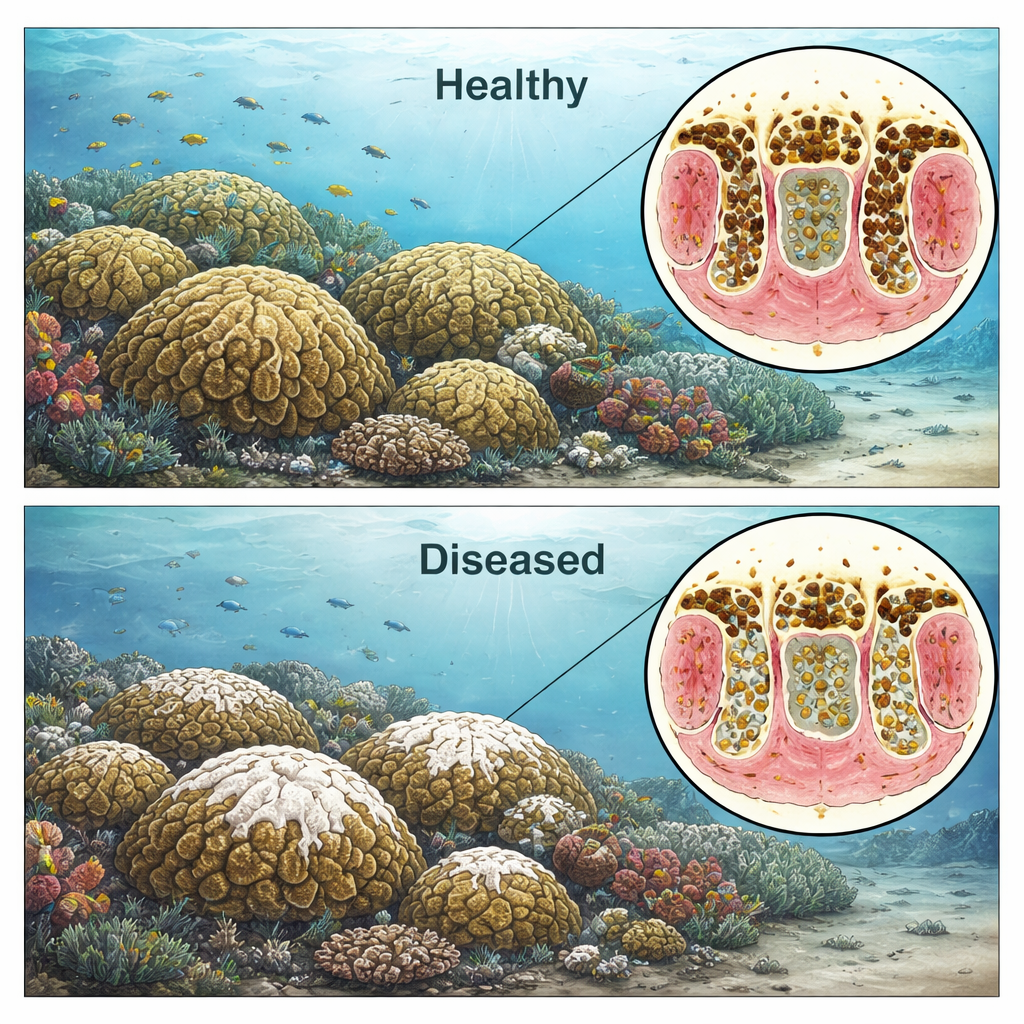
Measuring the slide from balance to breakdown
The team focused on four key features inside coral tissues: how large the algae were, how much the space around them (called a vacuole) had expanded, how often algae were being expelled from coral cells, and whether the layer of cells housing the algae was peeling away from the coral’s internal support structure. By calculating the ratio of algal cell size to vacuole size, they could quantify how tightly the partners remained connected. Using statistical models, they showed that this single ratio strongly predicted whether tissue came from a healthy-looking or diseased area. When the vacuole grew to more than about twice the area of the algal cell—meaning the algae were shrunken and surrounded by a large empty space—the odds that the coral was diseased rose sharply. In contrast, close contact between algae and host cells was linked to healthier tissue.
Runaway trouble in different coral and algae types
Not all corals, or their resident algae, responded the same way. Some highly vulnerable coral species, such as Colpophyllia natans, showed intense vacuole expansion, shrinking algae, and more algae being expelled, consistent with a severe breakdown in the partnership that could starve the coral and weaken its tissue. A more resistant species, Porites astreoides, showed different patterns, hinting that it may be better at recognizing and getting rid of problem algae before damage spirals out of control. When the team grouped samples by the dominant algal genus inside them—such as Cladocopium, Durusdinium, Breviolum, or Symbiodinium—they again found strong links between enlarged vacuoles, higher rates of algal expulsion, and diseased tissue for most algal groups. This suggests that which algae a coral hosts can shape how SCTLD unfolds at the cellular level. 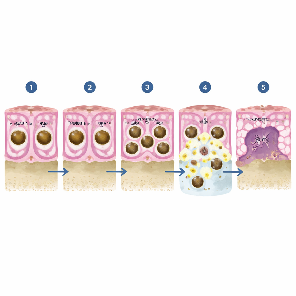
Signals from coral and algal genes
To connect what they saw under the microscope with deeper biological processes, the researchers paired tissue measurements with gene activity data from both corals and their algae. Certain coral genes involved in immune defense and in maintaining the structure of cell layers were more active when algae remained closely nestled in smaller vacuoles, hinting that a strong immune system and intact tissue scaffolding help keep the partnership stable. On the algal side, genes involved in stress management and in targeting viral genetic material for destruction were linked to healthier-looking cells and tighter coral–algae contact. These patterns support emerging evidence that SCTLD may involve viruses infecting the algae, and that both partners’ ability to manage stress and fight infection can influence whether tissues remain intact or begin to fall apart.
What this means for coral reefs
Taken together, the findings paint SCTLD not just as a disease of the coral animal, but as a runaway breakdown of the coral–algae partnership that begins inside individual cells. Once vacuoles swell and algae start to degrade and get expelled, the tissue that houses them weakens and pulls away, eventually leading to the dramatic peeling and loss of tissue seen on whole colonies. Differences among coral species and algal partners in how quickly and strongly this runaway process kicks in may explain why some corals succumb rapidly while others fare better. By turning subtle cellular changes into measurable indicators, this work provides new tools for diagnosing SCTLD, comparing outbreaks across regions, and ultimately helping managers target interventions that preserve the most resilient coral–algae partnerships.
Citation: Rossin, A.M., Beavers, K.M., Karrick, C.E. et al. Runaway coral-algal dysbiosis may be responsible for rapid coral tissue loss. Sci Rep 16, 6415 (2026). https://doi.org/10.1038/s41598-026-35666-4
Keywords: coral disease, stony coral tissue loss disease, coral-algal symbiosis, reef health, marine ecosystems