Clear Sky Science · en
Upright and supine assessment of pelvic floor muscle defects in women with and without prolapse
Why Pelvic Support Matters
Many women notice a feeling of heaviness, pressure, or bulging in the pelvis as they age or after childbirth, but the exact cause is often hidden deep inside the body. This study peeks under the surface using MRI scans to see how the muscles that support the pelvic organs hold up in different life stages—and what changes when pelvic organ prolapse develops. Understanding these hidden structures helps explain common symptoms and may guide better diagnosis and treatment in the future.
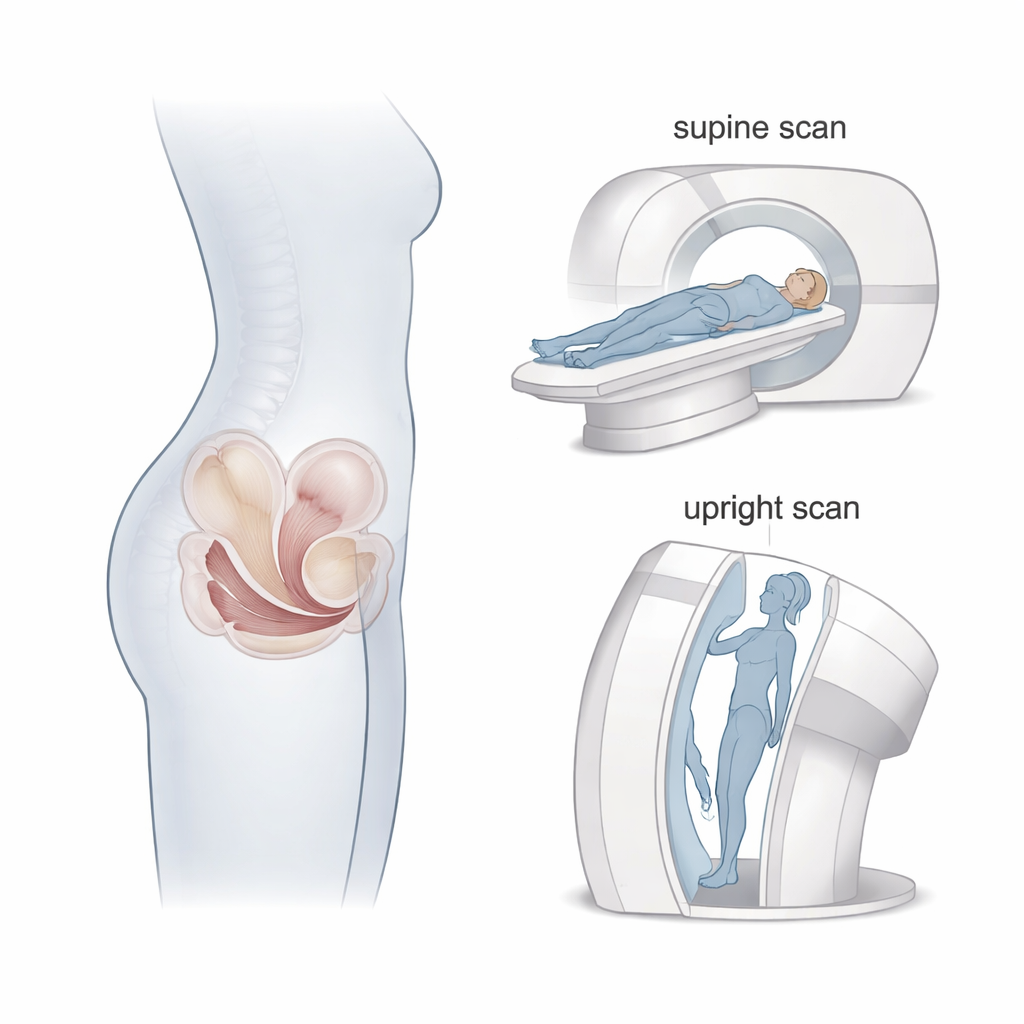
The Hidden Hammock Inside the Pelvis
At the base of the pelvis lies a sling of muscles that acts like a hammock, keeping the bladder, uterus, and bowel in place. Two key parts of this hammock—the pubococcygeus and the iliococcygeus—were the focus of this research. When sections of this hammock thin, bulge, or tear, organs can sag downward, leading to pelvic organ prolapse, incontinence, or problems with bowel control. The authors wanted to know how often such damage appears in women with and without prolapse, and whether body position during scanning—lying down or nearly upright—changes what doctors can see.
Studying Women Across Life Stages
The researchers enrolled 63 women and divided them into four groups: younger women who had never given birth; women who had given birth and were still before menopause; women who had given birth and were past menopause; and women with clear symptoms of pelvic organ prolapse. All participants underwent MRI scans in both a lying (supine) and almost standing (upright) position in a special tilting scanner. The team carefully graded muscle damage as none, minor, or major, and measured a key geometric feature called the iliococcygeus angle—the opening of the hammock—at several locations from front to back inside the pelvis.
Where Damage Shows Up—and Where It Doesn’t
Clear patterns emerged. Women who had never given birth showed no major damage in the pubococcygeus muscle, although a few already had small irregularities in the iliococcygeus portion of the hammock. Among women who had given birth, minor defects became more common, especially after menopause, and a small number showed major tears even without symptoms. In sharp contrast, most women with prolapse had major damage to the pubococcygeus muscle, and every prolapse patient had at least minor damage to the iliococcygeus, with many showing large bulges or hernias. These differences suggest that while small flaws in the pelvic hammock may be common, extensive breakdown is closely tied to prolapse.
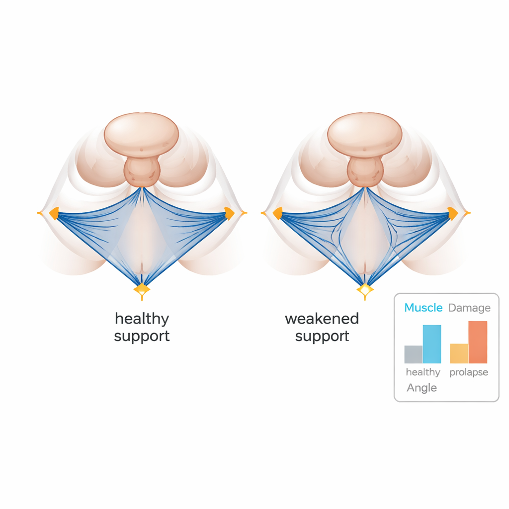
How the Pelvic Hammock Changes Shape
The shape of the muscle hammock itself also differed between groups. The iliococcygeus angle, which can be imagined as how wide the hammock opens across the pelvis, was much wider in women without prolapse and significantly sharper in those with prolapse, especially in regions under the vaginal canal and rectum. A wider angle in this study was interpreted as indicating better overall muscle support, while a sharper, more pinched angle marked a hammock that no longer effectively cradled the organs. These differences were easiest to detect in upright scans, when the weight of the organs pressed down in a more natural way, underscoring the value of imaging women in the position where their symptoms actually occur.
What This Means for Women’s Health
For a lay reader, the key message is that the pelvic floor is a working support system that can stretch, fray, and sometimes fail over time, particularly after childbirth and menopause. Minor wear and tear appears to be common and not always harmful, but large defects and a misshapen muscle hammock are strongly linked to prolapse. Upright MRI provides a more realistic picture of how well this hammock holds up under everyday gravity. Over time, such insights may help clinicians better predict who is at risk, tailor pessary or surgical treatments, and design exercises or therapies aimed at preserving this vital internal support.
Citation: de Alba Alvarez, I., Noort, F.v.d., Simonis, F.F. et al. Upright and supine assessment of pelvic floor muscle defects in women with and without prolapse. Sci Rep 16, 5600 (2026). https://doi.org/10.1038/s41598-026-35598-z
Keywords: pelvic organ prolapse, pelvic floor muscles, upright MRI, women's health, childbirth and menopause