Clear Sky Science · en
Regional ventilation–perfusion changes after endobronchial valve therapy assessed by single-photon emission computed tomography in adults with severe COPD
Why this matters for people with breathing troubles
For millions living with severe chronic obstructive pulmonary disease (COPD), even walking across a room can feel like climbing a mountain. Doctors now have a minimally invasive procedure that can collapse the most damaged parts of the lung using tiny one-way valves, sometimes easing breathing and boosting energy. This study asks a key question: can an advanced 3D scan called SPECT/CT show, in detail, how air and blood flow shift inside the lungs after this valve treatment, and could that eventually help pick which patients are most likely to benefit?
A closer look at damaged lungs
COPD destroys the tiny air sacs that exchange oxygen, leaving lung areas overinflated and inefficient. These unhealthy regions trap air and crowd out the healthier lung, making every breath harder. The valve procedure, known as bronchoscopic lung volume reduction, does not remove lung tissue but places small one-way valves into the airways of the worst lobe. Air can escape but not re-enter, allowing that lobe to shrink so the rest of the lung and the diaphragm can move more freely. Until now, doctors have mostly relied on CT scans and a catheter-based test to judge who should get valves, but those tools do not show in detail how air and blood are actually distributed within each lobe.
A new way to map air and blood flow
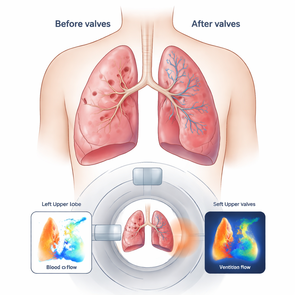
The researchers tested whether SPECT/CT—a scan that combines 3D X-ray images with tracers that track inhaled air and blood—could measure changes in lung function before and after valve placement. They enrolled six older adults with very severe emphysema and marked lung overinflation. Each patient received valves in a single, badly damaged lobe. Before and about three months after the procedure, the team performed SPECT/CT scans while patients inhaled a weakly radioactive mist to show ventilation and received an injection to show blood flow. Specialized software then divided the lungs into lobes and calculated what fraction of total air, blood, and volume each lobe contributed.
What changed inside the lungs
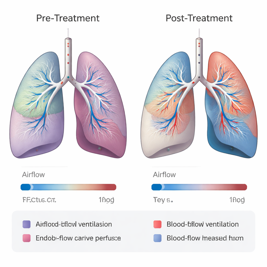
After valve treatment, the targeted lobe showed striking drops in function: its share of air flow fell by about two-thirds, blood flow by about three-quarters, and its volume shrank by roughly one-quarter. In other words, the valves successfully shut down and deflated the diseased region. At the same time, neighboring lobes on the same side of the chest picked up the slack. They gained air flow, blood flow, and volume, suggesting that both air and blood were being redirected to relatively healthier tissue. In contrast, lobes on the opposite side of the chest changed very little, indicating that the main reshuffling happened near the treated area. The SPECT/CT images clearly captured these shifts, lobe by lobe, in all six patients.
Body performance versus lung maps
Despite these dramatic internal changes, the improvements in whole-body measures were modest. On average, patients walked a bit farther in six minutes and had slightly better breathing tests, but the gains were small and varied from person to person. When the team compared changes seen on SPECT/CT with changes in walking distance and standard lung function, the relationships were weak. Some patients had large redistributions of air and blood flow without feeling much better, highlighting how factors like long-standing muscle weakness, heart health, or lingering lung stiffness can blunt the benefits of even well-placed valves.
What this could mean for future care
This early, small study suggests that SPECT/CT can act like a detailed weather map for the lungs, showing exactly how air and blood are rerouted after valve treatment. While it is too soon to use this scan to decide who should get valves, the results hint that, with larger studies and longer follow-up, such detailed imaging could help doctors choose the best target lobe, recognize patients unlikely to benefit, and perhaps extend similar mapping to other lung diseases. For people with severe COPD, that could eventually translate into more personalized treatment plans and a better chance that each invasive procedure truly pays off in easier breathing and improved daily life.
Citation: Karmali, D., Ghosh, P., Spottiswoode, B. et al. Regional ventilation–perfusion changes after endobronchial valve therapy assessed by single-photon emission computed tomography in adults with severe COPD. Sci Rep 16, 5153 (2026). https://doi.org/10.1038/s41598-026-35460-2
Keywords: COPD, endobronchial valves, lung imaging, ventilation perfusion, SPECT CT