Clear Sky Science · en
Introduction of the Ex/AxI ratio for optimized assessment of exophthalmos based on bulbar position parameters examined in a population-based MRI study
Why Eye Position Matters
Most of us take it for granted that our eyes sit neatly within their sockets. But when the eyeballs begin to bulge forward—known as exophthalmos—it can signal problems ranging from thyroid disease to tumors, and can threaten both vision and appearance. Doctors use simple rulers and mirrors to judge how far the eyes protrude, yet these readings can be skewed by natural differences in skull shape, body size, and eyeball length. This study introduces a more personalized way to judge eye bulging, using a new ratio called Ex/Axl, drawn from detailed MRI scans of a large slice of the German population.
Looking Closely at Eye Position
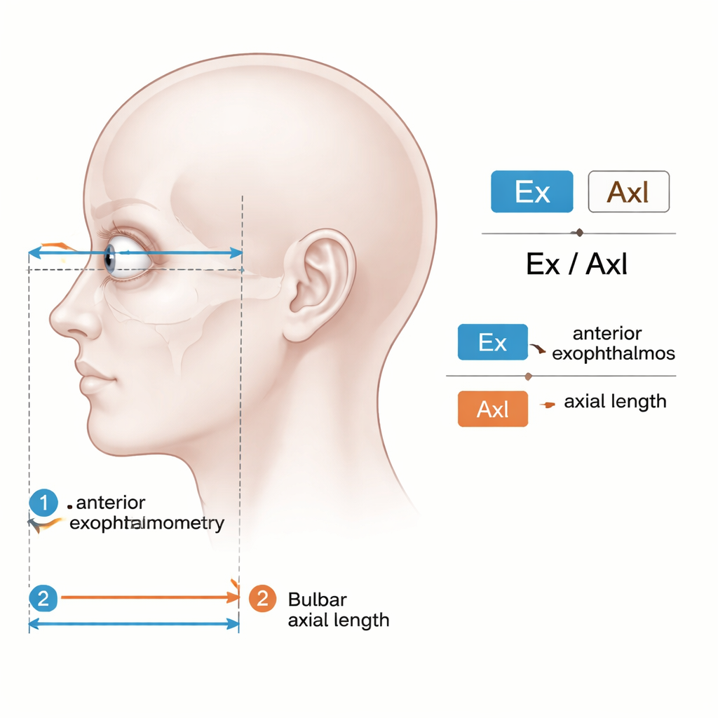
Traditional exophthalmometry focuses on how far the front of the eye sticks out relative to the bony rim of the eye socket. The authors argue that this “one-size-fits-all” measure ignores key individual differences, such as how long the eye itself is from front to back. A very long eyeball can look more prominent without any real disease, an effect sometimes seen in strong nearsightedness. To address this, the team measured both the eyeball’s length and its position in the orbit using high‑resolution MRI scans, which allow them to see not just the front of the eye, but also its back and surrounding tissues.
A Population-Based MRI Approach
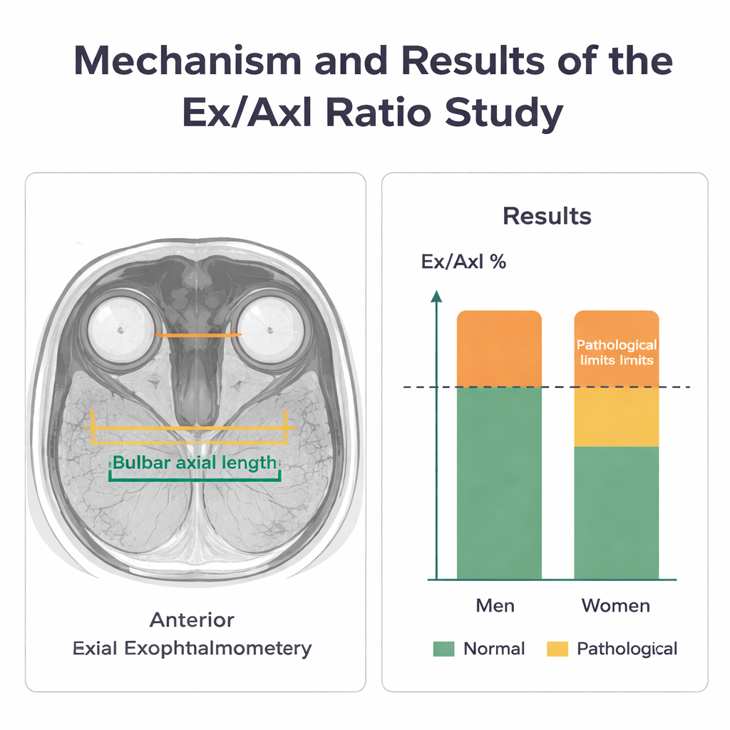
The researchers drew on over 6,700 adults participating in the long-running Study of Health in Pomerania. After excluding people who could not undergo MRI or whose images were too blurry, they analyzed scans from 1,926 participants aged 21 to 89. For each eye, they measured three distances: the axial length of the eyeball from front to back; the forward position of the cornea relative to a line drawn across the cheekbones (anterior exophthalmometry); and the depth of the back of the eye relative to that same line (posterior exophthalmometry). They then combined the first two into a single percentage: anterior protrusion divided by total eyeball length—the Ex/Axl ratio.
A New Ratio and Sex-Specific Limits
Because bodies differ, the team paid particular attention to sex, age, and body build. Men tended to have longer eyes and higher anterior protrusion values, while women showed slightly higher values when looking at the back of the eye. To define what should count as “normal,” the researchers created a reference group of healthier, average‑sized adults: over 30 years old, with no reported thyroid disease, no severe obesity, and moderate eyeball length. Within this group, they calculated the upper reference limits for their measurements. An Ex/Axl ratio above about 85% in men or 80% in women marked eyes that were unusually prominent for that person’s eyeball length and sex in this population, and therefore more likely to reflect a medically important bulging rather than just a large or elongated eye.
Linking Eyes to Body Build and Metabolism
The study went further by examining how eye position relates to overall body measurements and blood markers. Larger body weight, higher body‑mass index, and greater waist and hip circumference were all associated with higher Ex/Axl ratios in both men and women, suggesting that body build influences how full the orbit is and how far the eye sits forward. Certain blood fats and longer‑term blood sugar markers also showed links to the ratio, echoing hints from other research that metabolism, cholesterol, and thyroid function may interact with changes around the eye, even in people without overt thyroid eye disease.
What This Means for Patients
For patients and clinicians, the key message is that eye bulging should not be judged by a single millimeter cut‑off alone. By relating how far the eye protrudes to how long the eye itself is, and by using sex‑specific reference values drawn from almost two thousand MRI scans, the Ex/Axl ratio offers a way to distinguish harmless “big eyes” from bulging that may reflect disease. While MRI will remain a specialized tool, this work lays the groundwork for more personalized assessments of exophthalmos and for better tracking of treatments, such as surgery to relieve pressure in thyroid eye disease.
Citation: Lüdtke, L., Ittermann, T., Jürgens, C. et al. Introduction of the Ex/AxI ratio for optimized assessment of exophthalmos based on bulbar position parameters examined in a population-based MRI study. Sci Rep 16, 2599 (2026). https://doi.org/10.1038/s41598-026-35424-6
Keywords: exophthalmos, eye MRI, orbit anatomy, thyroid eye disease, ocular protrusion