Clear Sky Science · en
Dynamic thalamocortical functional connectivity disruptions in Parkinson’s disease with probable REM sleep behavior disorder
Why Nighttime Movements Matter
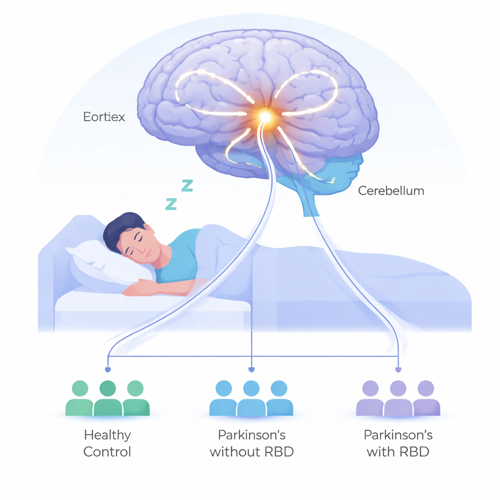
People with Parkinson’s disease often face more than tremors and stiffness. Many also act out their dreams while asleep—kicking, punching, or shouting in a condition called REM sleep behavior disorder (RBD). These episodes can injure patients and partners and may signal a faster-progressing form of Parkinson’s. This study asks a simple but important question: what is happening in the sleeping brain that turns quiet dreams into full‑body action?
Three Groups, One Big Question
The researchers compared three groups of volunteers: people with Parkinson’s and probable RBD, people with Parkinson’s but no RBD, and healthy adults. Everyone underwent brain scans while resting quietly in the MRI machine, along with detailed movement, mood, and thinking tests. By keeping age, disease duration, and medication levels similar between the Parkinson’s groups, the team could home in on what specifically distinguished those who acted out their dreams from those who did not.
Watching Brain Conversations in Motion
Instead of treating the brain as a static organ, the scientists focused on how communication between regions changes from moment to moment. They paid special attention to tiny hubs inside the thalamus—a deep relay station that passes information between the body, the outer brain surface (the cortex), and the cerebellum at the back of the brain. Using a technique called resting‑state functional MRI, they measured how strongly each thalamic hub’s activity rose and fell in sync with different parts of the brain over time, capturing the “fluctuations” in these connections rather than just their average strength.
A Sleep‑Linked Circuit Stands Out
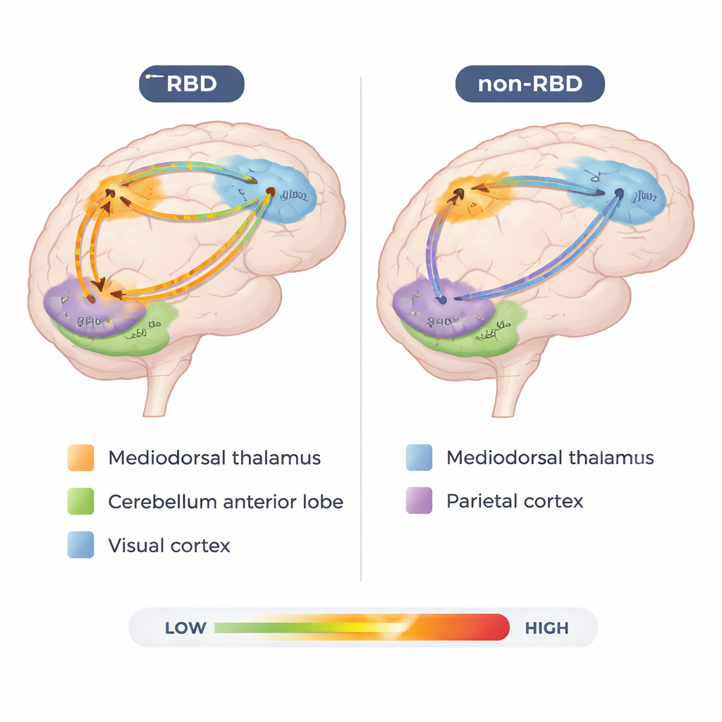
The most striking differences centered on a specific loop linking the mediodorsal part of the thalamus and the front portion of the cerebellum. In people with Parkinson’s and probable RBD, this circuit showed particularly large swings in connection strength, suggesting unstable communication. These fluctuations were not random: the more unstable this thalamus‑to‑cerebellum pathway was, the more severe a person’s dream‑enacting behaviors, as measured by a standard RBD questionnaire. That clear link between a single brain circuit and symptom severity points to a key pathway where sleep, movement control, and higher‑level planning may collide.
Different Parkinson’s, Different Brain Patterns
The group without RBD did not simply look “healthier.” Instead, they showed their own distinct pattern of altered connections. In particular, they had stronger and more variable links between another thalamic region, the pulvinar, and areas of the parietal cortex involved in attention and sensory integration. Meanwhile, people with RBD showed unique changes between the pulvinar and visual areas at the back of the brain, which may help explain their vivid, often disturbing dream imagery. Another sensory relay hub, the ventral posterolateral nucleus, was more tightly coupled with the cerebellum only in the RBD group, hinting at faulty filtering of body sensations during REM sleep when muscles should normally be paralyzed.
What This Means for Patients and Caregivers
Taken together, the findings suggest that acting out dreams in Parkinson’s is tied to instability in specific brain circuits rather than a simple worsening of overall disease. The thalamus, long viewed mainly as a relay station, emerges here as a dynamic control center whose shifting connections with the cerebellum and the cortex help determine whether dreams stay safely in the mind or spill out into the bedroom. If future, larger and longer‑term studies confirm these patterns, scans of these circuits could help doctors identify patients at higher risk, track how their disease is changing, and eventually guide targeted treatments to calm the nighttime storm while sparing waking function.
Citation: Tan, S., Zhang, Y., Niu, M. et al. Dynamic thalamocortical functional connectivity disruptions in Parkinson’s disease with probable REM sleep behavior disorder. Sci Rep 16, 4880 (2026). https://doi.org/10.1038/s41598-026-35415-7
Keywords: Parkinson’s disease, REM sleep behavior disorder, thalamocortical connectivity, resting-state fMRI, cerebellar circuits