Clear Sky Science · en
Non-invasive screening of alzheimer’s disease via label-free tri-spectral retinal imaging
Why the Eyes May Reveal Early Alzheimer’s
Alzheimer’s disease slowly damages the brain many years before memory problems become obvious, but current tests that look for these early changes are expensive, invasive, and not widely available. This study explores a simpler idea: because the retina at the back of the eye is a direct extension of the brain and can be photographed in seconds, could a regular eye exam—enhanced with smarter imaging—offer an easier way to spot Alzheimer’s at an earlier, more treatable stage? 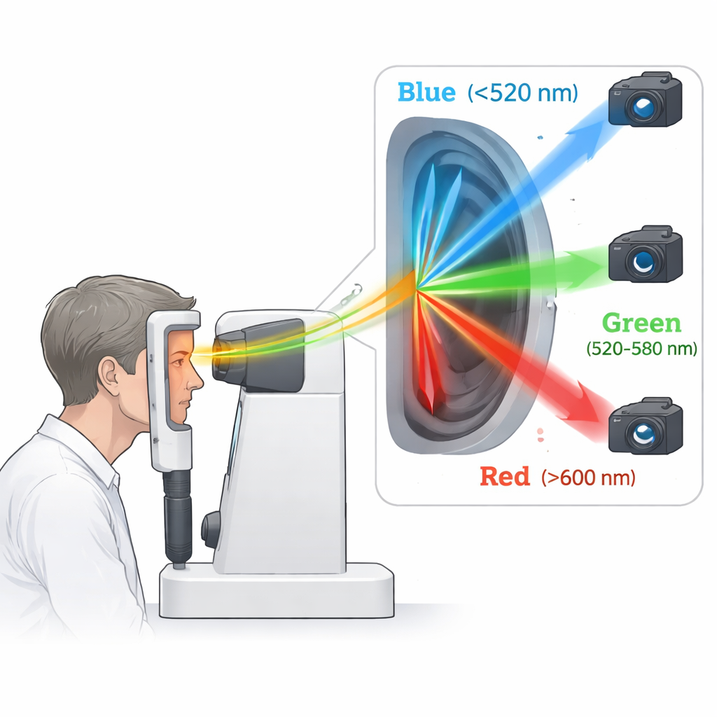
A New Way to Look at the Back of the Eye
The researchers built a compact add-on for a standard retinal camera, the same type often used in routine eye checkups. Instead of taking a single color photograph, their device splits the light reflected from the retina into three carefully chosen color bands: blue, green, and red. Each band is captured at the same moment by its own camera, ensuring sharp images without extra flashes or discomfort. This targeted approach improves how well subtle changes in the retina can be detected, especially in the shorter blue wavelengths, which earlier studies suggested are sensitive to Alzheimer’s-related changes.
Hidden Color Clues in the Retina
The team then tested this tri-spectral system in a clinical study including 38 people with Alzheimer’s disease, confirmed by brain scans or spinal fluid tests, and 28 healthy volunteers of similar age. After aligning all images so that key landmarks like the optic disc and fovea lined up pixel by pixel, they compared how much blue and green light was reflected from different parts of the retina. At first glance, the average images from patients and healthy subjects looked very similar. But when they calculated the ratio of blue to green light, a striking pattern appeared: the region between the central vision area (the fovea) and the optic disc showed a noticeably higher blue-to-green signal in people with Alzheimer’s. This difference, summarized by a performance measure called AUC of 0.74, suggests that color shifts in this nasal region of the retina carry useful information for distinguishing patients from healthy individuals. 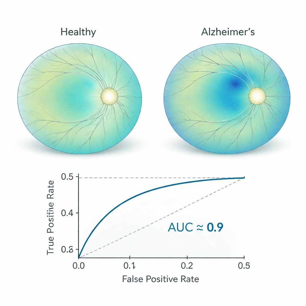
Teaching a Computer to Read the Signals
To turn these subtle optical fingerprints into a practical screening tool, the researchers trained a machine-learning model called XGBoost. Instead of relying only on simple ratios, the model used the raw blue, green, and red intensities from the most informative retinal region, along with age, sex, and basic eye-history information. It was trained on most of the eyes in the study and then tested on a separate group that the algorithm had never seen. On this independent test set, the model correctly separated Alzheimer’s and healthy eyes at a high level of accuracy, reaching an AUC of 0.91. Using an interpretability method known as SHAP, the authors showed that the blue-light measurements contributed most strongly to the model’s decisions, supporting the idea that Alzheimer’s-related chemistry in the retina affects how it scatters short-wavelength light.
What This Could Mean for Future Checkups
Because the tri-spectral module simply attaches to an existing fundus camera and requires only a single flash of light, it could in principle be built into routine eye exams without adding much time or discomfort for patients. Unlike brain scans or spinal fluid tests, this approach is non-invasive, relatively low-cost, and well suited to screening large numbers of people who may be at risk but not yet symptomatic. The authors emphasize that larger studies are needed and that this test would likely complement, not replace, established methods. Still, their findings suggest that carefully measuring how the retina reflects different colors of light—and letting transparent machine-learning tools interpret those patterns—may offer a practical new window into the earliest stages of Alzheimer’s disease, when interventions stand the best chance of altering the course of the illness.
Citation: Salajková, Z., Ciasca, G., Di Lorenzo, F. et al. Non-invasive screening of alzheimer’s disease via label-free tri-spectral retinal imaging. Sci Rep 16, 5083 (2026). https://doi.org/10.1038/s41598-026-35383-y
Keywords: Alzheimer’s disease, retinal imaging, early detection, non-invasive screening, machine learning