Clear Sky Science · en
Variability, asymmetry and sexual dimorphism in craniofacial anomalies in Loeys-Dietz syndrome 2: geometric morphometric analysis in mice
Why this rare disorder matters to faces and health
Loeys-Dietz syndrome is a rare inherited condition best known for dangerous bulges in the body’s largest artery, but it also alters how the skull and jaw grow. These changes can affect breathing, chewing, appearance, and quality of life. This study uses a specially bred mouse that carries the same gene change as people with one form of Loeys-Dietz syndrome to show when and how facial bones go off course, why the severity varies so much, and why girls and women may be more strongly affected. 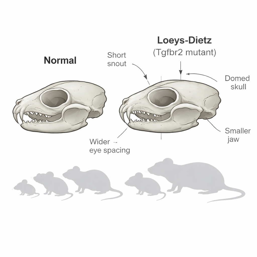
A rare disease with a visible face
Loeys-Dietz syndrome arises from faults in a cell-to-cell messaging system known as TGF-beta signaling. One of the most severe subtypes, called type 2, is caused by changes in a receptor gene named TGFBR2. Patients with this subtype can show a broad range of facial differences: wide-set eyes, a short or recessed jaw, a high arched palate, dental crowding, and sometimes premature fusion of skull seams. Earlier clinical work from the same group showed that these traits vary greatly from one patient to another and that people with type 2 tend to have the most pronounced and diverse facial changes. Dentists and surgeons who care for these patients often face complex treatment decisions, yet it has been hard to study how and when these features emerge because young children are rarely exposed to 3D skull scans.
Following skull growth in a living model
To fill this gap, the researchers turned to a knock-in mouse that carries the exact Tgfbr2 mutation found in human type 2 patients. Unlike earlier mouse models where the gene was completely shut off in certain cells, these mice more closely mimic the human situation, carrying one normal and one altered copy of the gene on a uniform genetic background. The team scanned the skulls of 84 mice at four stages roughly corresponding to early infancy, late childhood, young adulthood, and mature adulthood in humans. Using high-resolution micro–CT and 3D geometric morphometrics—a way of capturing shape with dozens of anatomical landmarks—they compared overall skull and jaw form, measured lengths and angles, and quantified left–right differences.
Early, variable, and asymmetric changes in skull and jaw
The mutant mice already showed clearly different skull shapes by two weeks of age, suggesting that facial changes are present around birth rather than emerging only during growth. Compared with healthy littermates, affected mice had shorter front portions of the skull, wider spacing between the eye sockets, smaller mandibles, and a domed skull outline. The joints and hinges of the jaw were especially altered: the condyle at the back of the jaw grew irregularly and often became mushroom-shaped, and a bony projection called the coronoid process stretched backward. Computerized overlays and heat maps confirmed that these differences were not uniform. Instead, each mutant skull deviated from the norm in its own way and frequently showed marked left–right asymmetry in both the upper and lower jaws. This mirrors the wide range of facial appearances and jaw joint problems observed in human patients. 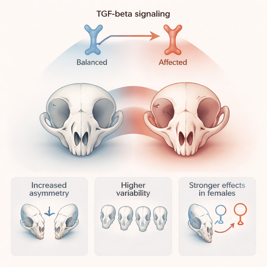
Sex differences that echo from mice to people
When the team examined individual features, some stood out as more common or more severe in female mice. These included cranial doming, pronounced bending of the nasal region, greater shortening of the skull base, and stronger differences between the two sides of the jaw. Statistical tests on shape data suggested that, although overall skull form did not split neatly by sex, females tended to occupy more extreme positions in the range of possible shapes. Prompted by this, the researchers revisited clinical records and 3D scans from 26 people with type 2 Loeys-Dietz syndrome. They found hints of the same trend: women more often had flatter midfaces, smaller jaws, greater mismatch between the upper and lower jaws, and more obvious nasal deviation linked to underlying bone asymmetry rather than just cartilage.
What this means for patients and future care
By showing that a single TGFBR2 mutation can produce early, highly variable, and often asymmetric changes in skull and jaw growth—even in genetically uniform mice—this work suggests that much of the facial diversity in Loeys-Dietz syndrome is built into how the mutation disrupts bone development, not only into background genes or environment. The close parallels between the mouse model and human patients, including possible stronger effects in females, make this system a powerful tool for dissecting the biology behind craniofacial anomalies and for testing new treatments. In the future, better understanding of these facial changes may help doctors predict which patients face higher risks, plan safer surgeries, and improve both function and appearance for people living with this rare but impactful condition.
Citation: Devine, K.R., Lynn, S., Jani, P. et al. Variability, asymmetry and sexual dimorphism in craniofacial anomalies in Loeys-Dietz syndrome 2: geometric morphometric analysis in mice. Sci Rep 16, 2185 (2026). https://doi.org/10.1038/s41598-026-35325-8
Keywords: Loeys-Dietz syndrome, craniofacial development, mouse model, facial asymmetry, sex differences