Clear Sky Science · en
Artificial intelligence classification of rectal neoplasia by endoscopic fluorescence perfusion analysis
Why this matters for patients and doctors
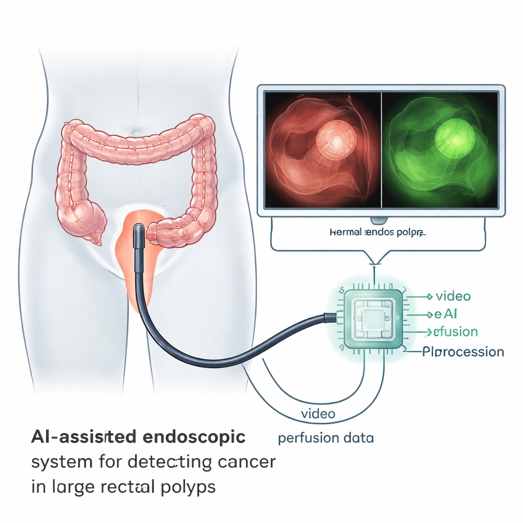
For people with large rectal polyps, one of the biggest questions is whether the growth is harmless or already turning into cancer. Today, doctors often cannot be sure until after they remove the entire lesion, which may mean bigger operations than necessary or delays in treatment. This study explores whether a smart imaging technique, combined with artificial intelligence, can spot hidden cancer during the procedure itself by watching how blood flows through the tissue.
Listening to how a tumor is fed
Cancers do not grow like normal tissue. They stimulate the formation of new, abnormal blood vessels that leak and branch in a disorganized way. These changes create distinctive patterns in how blood, and injected dyes, wash in and out of a tumor. The researchers used a dye called indocyanine green, which glows under near‑infrared light, and recorded short videos during endoscopic surgery in patients with large rectal polyps and early rectal cancers. By following the brightness of the glow over several minutes, they could capture a kind of “perfusion fingerprint” for both suspicious and healthy areas inside the same patient.
Turning glow patterns into data
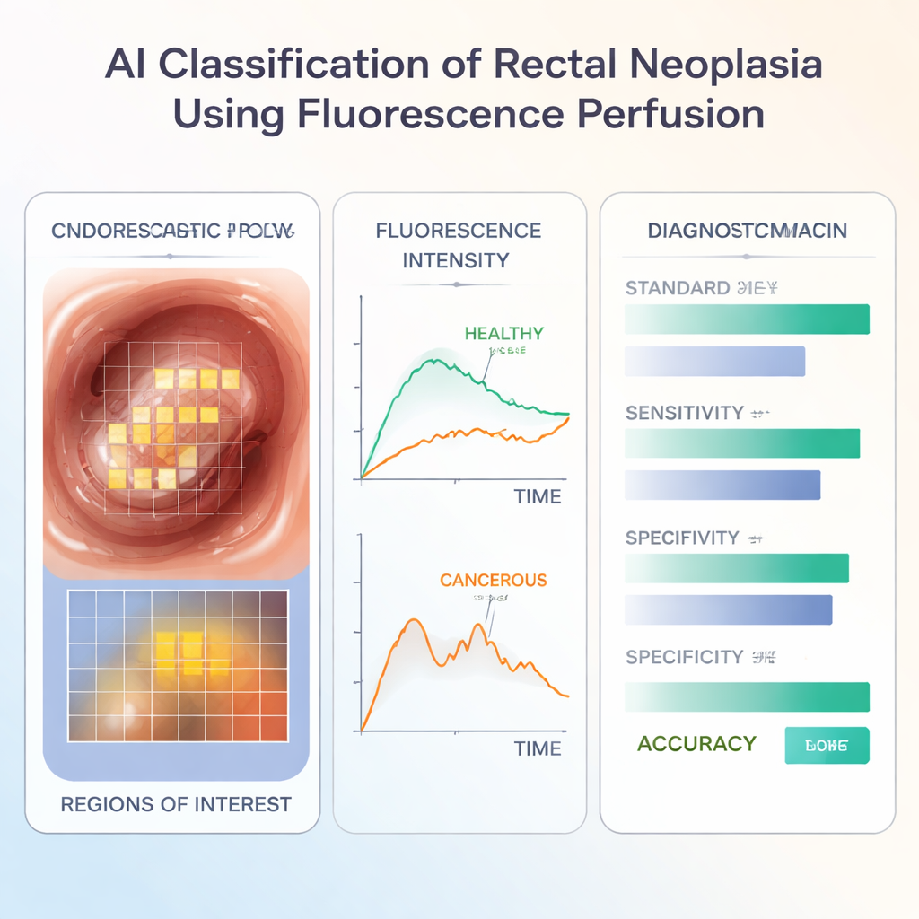
Each video was analyzed by custom software that divided the visible region of the bowel wall into a grid of tiny squares and tracked them over time, even as the camera and tissue moved. For every square, the program measured how bright the fluorescence became, how quickly it peaked, and how fast it faded. It then cleaned and normalized these curves so they could be compared directly. From these time traces, the team extracted simple numerical features such as the maximum signal and the drop in signal at specific times after the peak. They also looked at how uneven these values were across the abnormal region, using a statistic that captures variation within the tumor compared with nearby healthy tissue.
Training the artificial intelligence
The group studied 190 videos from 182 patients treated at six hospitals in four countries; about three in five patients ultimately had cancer confirmed under the microscope. They trained a machine‑learning model (an XGBoost classifier) to learn the difference between benign and cancerous lesions based only on the dye‑flow features, without looking at the normal color images. When used on new cases, the model correctly identified cancer in a substantial majority of patients, performing on par with or slightly better than many standard tools used in practice, such as endoscopic biopsies, pre‑operative MRI scans, and the expert surgeon’s visual impression.
Adding real‑world clinical clues
In real life, doctors rarely rely on a single test. The researchers therefore combined the AI’s output with information that is already available: MRI reports and the operating surgeon’s judgment. When these were fed into the same computer pipeline, the ability to detect cancer improved, especially in correctly ruling out disease in benign lesions. In the best scenario, the combined system detected about 86% of cancers while avoiding false alarms in roughly 71% of non‑cancer cases. The approach also worked reasonably well in subgroups of patients more typical of early, locally removable disease.
What this could mean for future care
The study shows that cancers in large rectal polyps leave a detectable signature in how blood and dye flow through them, and that this signature can be picked up automatically by AI. While the work so far has been done on recorded videos and still needs to be proven in real‑time clinical trials, it points toward a future in which a colonoscopist could be warned, during the procedure, that a seemingly harmless polyp likely hides an invasive cancer. That information could guide where to biopsy, whether to remove the lesion locally, or whether to refer the patient for more extensive surgery, reducing both missed cancers and unnecessary major operations.
Citation: Boland, P.A., MacAonghusa, P., Singaravelu, A. et al. Artificial intelligence classification of rectal neoplasia by endoscopic fluorescence perfusion analysis. Sci Rep 16, 4761 (2026). https://doi.org/10.1038/s41598-026-35233-x
Keywords: rectal cancer, endoscopic imaging, fluorescence perfusion, artificial intelligence, machine learning