Clear Sky Science · en
Evaluation of an ultrasound-guided alveolar recruitment technique with incremental PEEP in dogs: a clinical study
Why sleepy dogs’ lungs matter
When dogs go under general anesthesia for surgery or imaging, their lungs can quietly misbehave. Air sacs may collapse, making it harder for oxygen to reach the bloodstream—a problem called atelectasis. This study asks a timely question for modern veterinary medicine: can vets use a simple bedside ultrasound exam, together with a gentle pressure-based breathing technique, to spot and reverse these hidden lung collapses in real time, without invasive tests or complex machines?
Hidden lung trouble during routine anesthesia
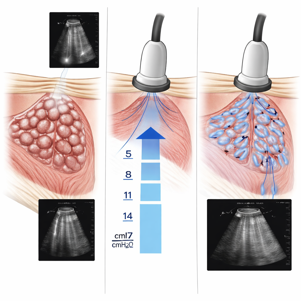
Atelectasis happens when tiny air sacs in the lungs, called alveoli, deflate or stick shut. During anesthesia, muscles relax, pets breathe differently, and they often receive pure oxygen. All of this can promote collapse in parts of the lung, even in otherwise healthy animals. Until recently, veterinarians had limited tools to detect these changes during the procedure. Traditional imaging like CT scans is expensive, slow, and not practical in the operating room. Lung ultrasound, by contrast, is portable, quick, and shows characteristic patterns—such as vertical bright lines or small solid-looking patches—that hint at collapsed regions.
Using sound waves to map problem areas
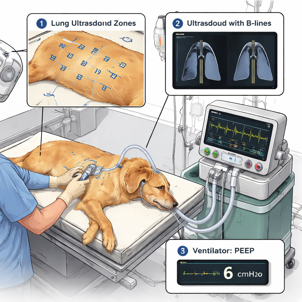
In this clinical study, 36 client-owned dogs were anesthetized for routine procedures. Some had surgery and were placed on a mechanical ventilator, while others underwent diagnostic imaging and continued to breathe on their own. Before anesthesia, all dogs received a structured lung ultrasound exam, with each side of the chest divided into nine regions. The researchers created a “lung ultrasound atelectasis score” (LUAS) that graded each region from normal to severely collapsed based on how many ultrasound signs of fluid or consolidation were seen. After the procedure, dogs were scanned again; if enough abnormal regions were found, the team assumed atelectasis was present and moved on to a targeted breathing intervention.
Gently reopening collapsed air sacs
The intervention was an “alveolar recruitment maneuver,” which means carefully using the ventilator to push air into the lungs with slightly higher pressure at the end of each breath, known as positive end-expiratory pressure (PEEP). The team started at a modest PEEP level and then increased it stepwise every two minutes, while repeatedly looking at the worst-affected lung region on ultrasound. The goal was to keep raising PEEP only until the ultrasound score in that region improved to near-normal, signaling that the collapsed air sacs had reopened. Throughout this process, the researchers closely monitored heart rate, blood pressure, breathing pressures, and how easily the lungs expanded, to make sure the maneuver remained safe.
What the scans and monitors revealed
The findings were striking: about 83% of the dogs showed ultrasound signs of atelectasis after anesthesia—much higher than older estimates based on other imaging methods. Dogs on mechanical ventilators tended to have more severe collapse and needed higher PEEP levels to fix it than dogs that were breathing on their own. As PEEP was increased, ultrasound scores steadily dropped toward normal, and after the maneuver was completed and the PEEP was turned back down to zero, lung compliance improved and the pressure needed to give each breath fell. Importantly, despite brief rises in heart rate at higher PEEP levels, blood pressure and overall heart output stayed within safe limits. A follow-up ultrasound 15 minutes after the breathing tube was removed showed that the improvement in lung appearance was maintained.
What this means for pet patients
For pet owners, the message is reassuring: even though lung collapse during anesthesia is common, it does not have to go unnoticed or untreated. This study shows that veterinarians can use a quick, non-invasive ultrasound exam to detect areas of collapse and then guide a tailored breathing strategy that reopens the affected lung regions while keeping circulation stable. In plain terms, sound waves and smart ventilator settings together give anesthetized dogs a better chance at maintaining healthy lungs before, during, and after routine procedures.
Citation: Di Franco, C., Boysen, S., Buonamici, B. et al. Evaluation of an ultrasound-guided alveolar recruitment technique with incremental PEEP in dogs: a clinical study. Sci Rep 16, 4830 (2026). https://doi.org/10.1038/s41598-026-35202-4
Keywords: canine anesthesia, lung ultrasound, atelectasis, ventilation, alveolar recruitment