Clear Sky Science · en
Hemi-ellipsoid formula enables accurate assessment of carotid plaque volume and atherosclerotic burden
Why the shape of artery plaque matters
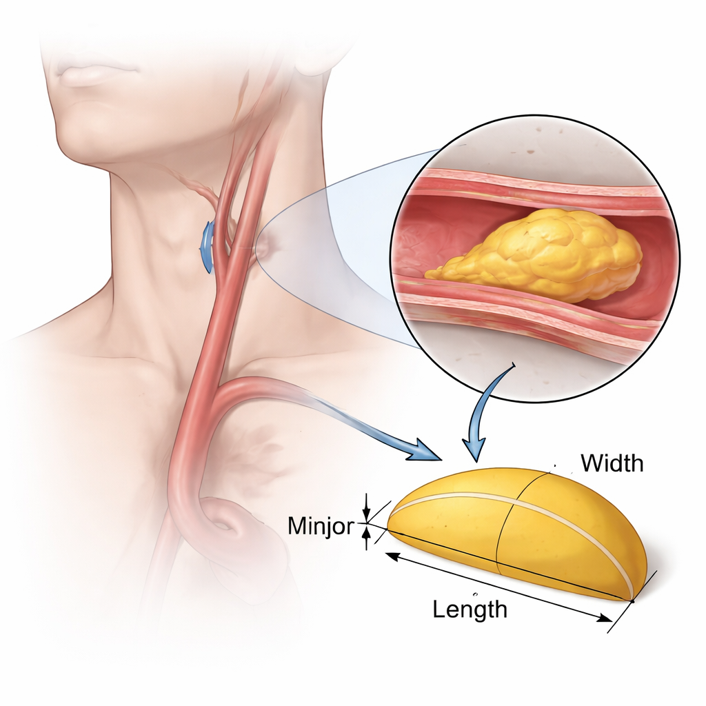
Stroke and heart attacks often begin silently, with fatty "plaques" slowly building up inside neck arteries that feed the brain. Doctors usually judge how dangerous these plaques are by how much they narrow the vessel in a single slice or picture. But plaques are three-dimensional bulges that grow in length, width, and height over time. This study shows that treating each plaque like a simple three‑dimensional shape—a half of an elongated sphere—can give a more accurate picture of its true size and growth, using the same routine ultrasound scans already done in clinics.
Turning a complex lump into a simple shape
Carotid plaques form inside tube‑shaped arteries and protrude into the blood channel in irregular ways. The researchers asked a practical question: can we logically approximate these messy shapes as a single, smooth "hemi‑ellipsoid"—like half of a stretched ball—so that their volume can be computed with a straightforward formula? Using standard carotid ultrasound images taken from the side and across the vessel, they measured three things: how far the plaque runs along the vessel, how far it wraps around the inside wall, and how far it sticks into the lumen on average. Plugging these three numbers into a hemi‑ellipsoid volume equation provides a quick estimate of how big the plaque really is in three dimensions.
Testing the idea with model plaques
To check whether this shortcut is trustworthy, the team first built mathematical “toy” plaques from combinations of hemispheres and half‑ellipsoids laid out on a flat surface. For these idealized shapes, they could calculate the exact volume and then compare it with the volume predicted by the hemi‑ellipsoid formula using only the total length, width, and thickness. For plaques with smooth or evenly thick surfaces, the shortcut matched the true volume very closely. When they made highly uneven shapes, the method initially overestimated volume if it used only the thickest part. Replacing the single maximum thickness with the average of the thickest and thinnest points brought the estimated volumes back in line with the true ones. The group then repeated this test with computer‑generated 3D plaque models inside a virtual artery and again found near‑perfect agreement.
From lab models to real patients
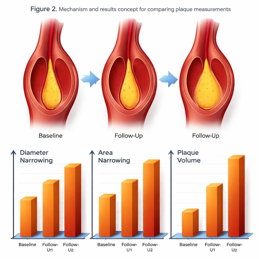
The researchers next asked whether this volume‑based approach would change how plaque progression looks in real people. They analyzed ultrasound scans from 115 stroke patients who were followed for more than seven years, tracking 373 carotid plaques. For each plaque, they calculated three measures at the start and at follow‑up: the traditional one‑dimensional percentage narrowing of the vessel, the two‑dimensional percentage loss of cross‑sectional area, and the new three‑dimensional plaque volume using the hemi‑ellipsoid formula. While diameter narrowing and area narrowing rose only modestly over time, plaque volume almost doubled. Many plaques even appeared to "improve" when judged by diameter or area alone, yet still grew in total volume when all three dimensions were accounted for.
Seeing total disease burden more clearly
Because most patients had several plaques, the team also summed all plaques for each person to estimate total atherosclerotic burden. By this more global view, no patient showed a true reduction in total plaque volume over the years, even though some seemed better when only narrowing in a single slice was considered. Statistical analyses that linked plaque severity to common cardiovascular risk factors such as age, blood sugar, and cholesterol also fit better when plaque volume was used instead of diameter or area. This suggests that three‑dimensional volume not only tracks growth more faithfully, but also mirrors the underlying biology of artery disease more closely.
What this means for everyday care
For patients and clinicians, the key message is that how much a plaque narrows an artery in one image does not tell the whole story. A plaque can grow longer and thicker without dramatically changing the measured percent narrowing, giving a false sense of stability. By treating each plaque as a simple half‑ellipsoid and using readily obtained ultrasound measurements, doctors can estimate its true volume and follow how that volume changes over time. This work suggests that such volume tracking offers a more reliable way to judge stroke‑related plaque burden and to monitor the impact of treatments aimed at slowing or reversing artery disease—without needing complex 3D imaging in everyday practice.
Citation: Kim, J., Jeong, T. & Kim, J. Hemi-ellipsoid formula enables accurate assessment of carotid plaque volume and atherosclerotic burden. Sci Rep 16, 5138 (2026). https://doi.org/10.1038/s41598-026-35182-5
Keywords: carotid plaque, atherosclerosis, ultrasound imaging, plaque volume, stroke prevention