Clear Sky Science · en
Impact of meningioma and glioma on whole-brain dynamics
Why brain tumors affect more than just one spot
Brain tumors are often pictured as isolated lumps that press on nearby tissue. But our thoughts, movements, and emotions rely on signals that ripple across the entire brain. This study asks a deceptively simple question with far-reaching implications: do common brain tumors quietly reshape the way the whole brain communicates, even far from the visible lesion—and if so, does this differ between two major tumor types, meningiomas and gliomas?
Two common tumors with very different behavior
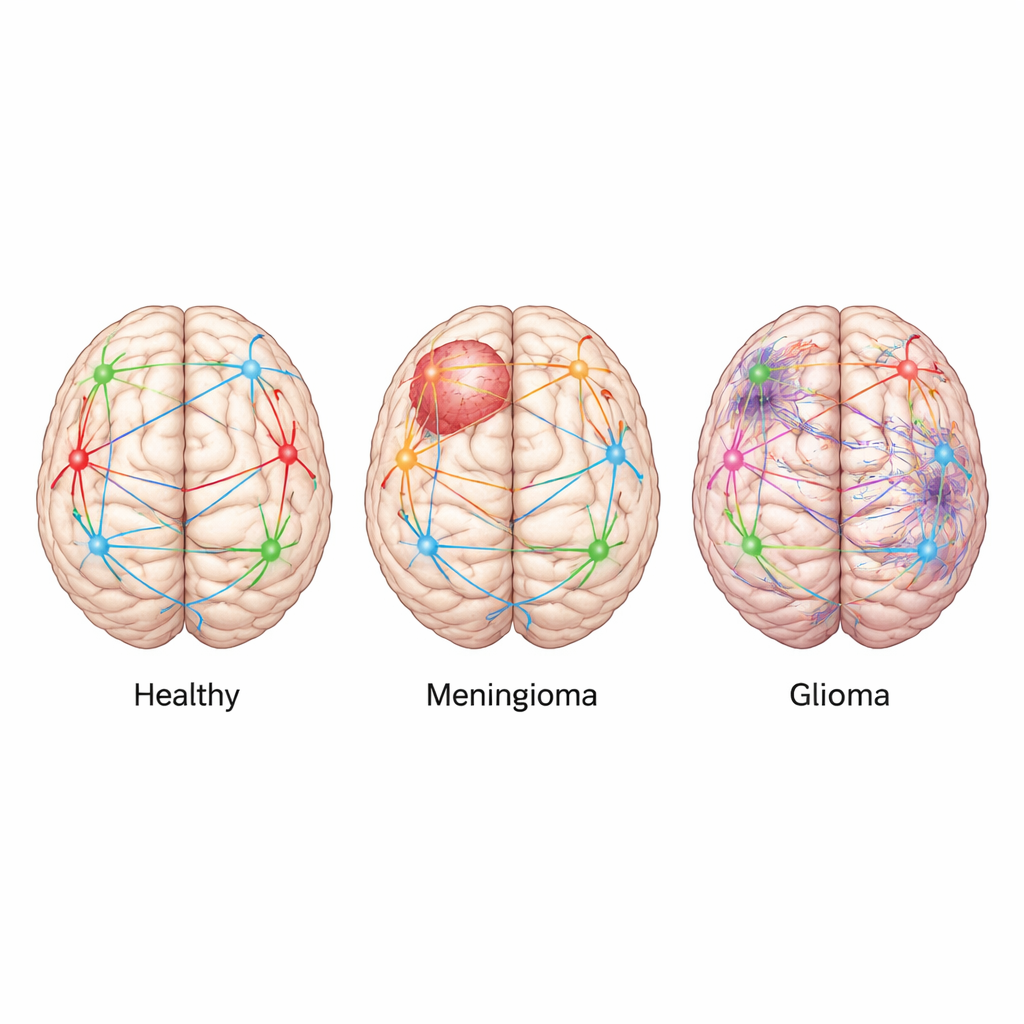
Meningiomas and gliomas are among the most frequent brain tumors, but they behave quite differently. Meningiomas usually grow from the brain’s protective membranes and tend to push on the brain from the outside, often remaining well defined and slow growing. Gliomas, in contrast, arise from supporting brain cells and infiltrate the surrounding tissue, weaving themselves into the brain’s wiring. To explore how these distinct growth patterns affect brain function, the researchers drew on MRI scans from 34 adults: 10 healthy volunteers, 10 people with gliomas, and 14 with meningiomas, all scanned before surgery while simply lying still in the scanner.
Measuring how brain signals ripple and flex
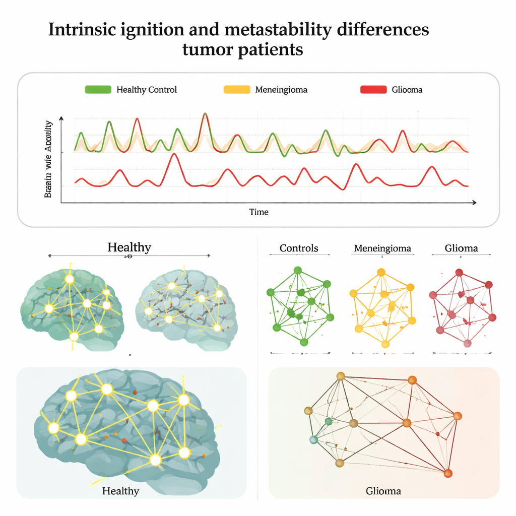
Instead of looking only at where the tumors sat, the team focused on how activity unfolded over time across the whole brain. They used resting-state functional MRI, which tracks slow changes in blood flow as a stand-in for neural activity, and applied a computational approach called the Intrinsic Ignition Framework. In simple terms, they asked two questions: how well can a local burst of activity in one region "ignite" wider communication across the brain, and how flexibly does the brain shift between more synchronized and more independent states? They called the first property "intrinsic ignition" and the second "metastability," and calculated both for each person and for specific brain regions and networks.
Gliomas disturb the whole network, meningiomas mainly near the tumor
When comparing groups, a stark pattern emerged. People with gliomas showed clearly reduced ignition and metastability compared with healthy volunteers, meaning their brains were less able to broadcast local signals and less flexible in coordinating activity over time. These disruptions appeared even in regions that looked tumor-free on conventional scans, consistent with the invasive nature of gliomas, which can send microscopic tendrils far from the main mass. In contrast, meningioma patients showed overall values much closer to healthy controls. Noticeable changes appeared mainly in regions where the tumor occupied more than about one third of the area, especially for ignition, suggesting that compression can blunt a region’s ability to drive communication while leaving much of the broader network architecture intact until the load becomes substantial.
Hidden network changes in key brain systems
The researchers then zoomed out to well-known resting-state networks, such as those involved in vision, movement, attention, and daydreaming (the "default mode" network). In healthy brains, metastability was strongly coordinated across these networks, and ignition and metastability tended to rise and fall together. Meningioma patients showed only a mild weakening of these relationships. Glioma patients, however, displayed markedly disrupted patterns: correlations between networks were fractured, and the usual coupling between how strongly regions ignite and how flexibly they synchronize was much weaker. Importantly, when the team linked these brain measures to performance on a computerized attention test, healthy volunteers with higher ignition in key networks responded faster. Tumor patients, despite achieving similar test scores overall, no longer showed this neat brain–behavior link, suggesting that their brains were relying on less efficient, compensatory routes.
What this means for patients and future care
Taken together, the findings support a simple but powerful message for non-specialists: not all brain tumors disturb the brain in the same way. Meningiomas, especially the mostly benign, slow-growing cases in this study, tend to cause local mechanical problems that only gradually spill over into wider communication issues. Gliomas, by contrast, act more like a disease of the brain’s wiring itself, quietly degrading communication patterns across distant regions. The study also shows that sophisticated measures of how signals ignite and fluctuate across the brain can reveal network damage even when routine tests look normal. In the future, such "dynamic fingerprints" may help doctors track how tumors disrupt information flow, tailor treatments, and monitor recovery beyond what scans of anatomy alone can show.
Citation: Juncà, A., Escrichs, A., Martín, I. et al. Impact of meningioma and glioma on whole-brain dynamics. Sci Rep 16, 5032 (2026). https://doi.org/10.1038/s41598-026-35140-1
Keywords: brain tumors, glioma, meningioma, brain networks, functional MRI