Clear Sky Science · en
Distal radius morphometry of volar curvature along with scaphoid and lunate facet inclinations and ulnar variance in the Anatolian population
Why Wrist Bone Shape Matters
Breaking a wrist is one of the most common bone injuries in adults, and many of these fractures are repaired from the palm side of the forearm using metal plates and screws. Yet those plates are usually sold in just a few “standard” shapes. This study asks a simple but important question: how well do those one‑size‑fits‑all plates actually match the true shape of the wrist bone in a real population—in this case, people from Anatolia (modern‑day Turkey)? The answer could affect how well fractures heal, how comfortably the wrist moves, and whether tendons are irritated by poorly fitting hardware.
The Hidden Curves of the Wrist Bone
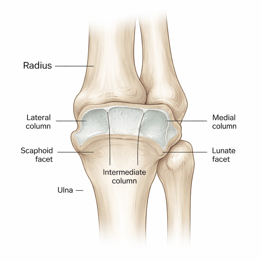
The research focuses on the far end of the radius, the larger of the two forearm bones, right where it meets the small wrist bones. On the palm (volar) side, this region is not flat; it has a gentle forward curve and two sloping joint surfaces that cradle neighboring wrist bones called the scaphoid and lunate. Surgeons rely on this surface when they attach plates to stabilize fractures. If a plate does not follow these curves, it can leave parts of the joint under‑supported or allow sharp edges to rub on nearby tendons. The team set out to measure these curves and slopes in careful detail, looking specifically at the bend of the front surface, the tilt of the scaphoid and lunate contact areas, the gap between the radius and the neighboring ulna, and the overall width of the volar surface.
Three‑Dimensional Scans Instead of Guesswork
To capture the true shape of the bone, the researchers analyzed three‑dimensional CT scans of 103 healthy wrists from adults aged 19 to 67. They excluded any scans with previous fractures or joint disease so that only normal anatomy was studied. Using specialized software, they built 3D models of each radius and defined standard planes that passed through key landmarks, such as the centers of the scaphoid and lunate contact areas. On these planes, they measured how strongly the front surface curved, one and two centimeters above the rim of the joint, and how steeply the scaphoid and lunate facets were tilted. They also measured how far the end of the ulna sat above or below the end of the radius—a value known as ulnar variance, which can alter how loads are shared across the wrist.
Distinct Patterns by Sex, But Not by Side
The results showed clear patterns. Men tended to have a more pronounced forward curve of the volar surface in all measured regions compared with women, meaning that their distal radius bends more strongly toward the palm. The width of the volar surface was about 26.5 millimeters on average, slightly wider in men but without large differences by age or by right versus left wrist. The tilt of the scaphoid and lunate facets also varied: on average, the scaphoid facet sloped forward, the lunate facet was close to flat or slightly backward, and the angle between them (the interfacet angle) was larger in men. Ulnar variance averaged about two millimeters negative, meaning the ulna usually ended a bit shorter than the radius, and this value tended to increase slightly with age. Importantly, no meaningful differences were found between right and left wrists, suggesting that the opposite, uninjured wrist can serve as a reliable template when planning surgery.
Why Shape Mismatch Can Cause Trouble
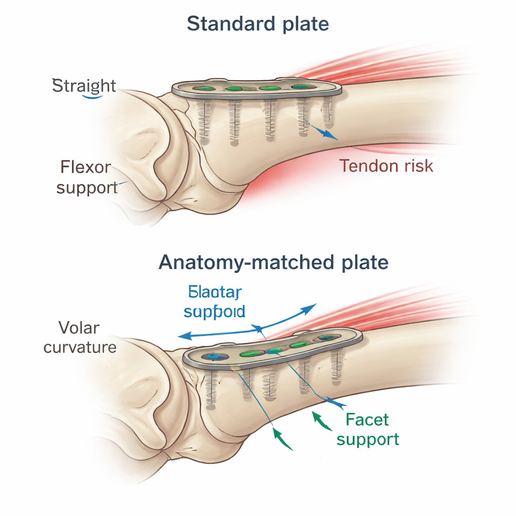
These measurements are more than academic. If a pre‑shaped plate is flatter than the actual bone, it may not fully support the forward‑projecting rim that holds the lunate, allowing that rim to shift and the wrist bones to slide forward over time. A plate that sits too far toward the joint can also encroach on the space of the flexor tendons that bend the fingers and thumb, increasing the risk of irritation or even tendon rupture. The study shows that curvature and facet angles vary with sex and age, and that many individuals diverge from the “average” bone used to design standard implants. As a result, a plate that fits one patient well may fit another quite poorly, even if both have similar fractures.
What This Means for Patients and Surgeons
For a lay reader, the takeaway is that the fine details of wrist bone shape matter for how well a fracture repair will perform in daily life. This work provides a detailed, three‑dimensional map of the volar radius in an Anatolian population, highlighting differences linked to sex and age and tying them to known surgical risks. Rather than relying on a single measurement or a generic plate, the authors argue that surgeons should consider several features together—the front‑to‑back curve, the slopes of the scaphoid and lunate facets, and the relative length of the ulna—when choosing and positioning implants. While fully custom plates are still uncommon, using 3D imaging and population‑based reference data can help tailor implant choice and placement, potentially leading to better wrist motion, fewer complications, and more durable repairs after a break.
Citation: İsmailoğlu, P., Nalbantoğlu, U., Tok, O. et al. Distal radius morphometry of volar curvature along with scaphoid and lunate facet inclinations and ulnar variance in the Anatolian population. Sci Rep 16, 4946 (2026). https://doi.org/10.1038/s41598-026-35123-2
Keywords: distal radius fractures, wrist anatomy, volar plate fixation, orthopedic implants, computed tomography